

Disseminated Pyomyositis Due to Burkholderia cepacia: A Case Report
- PMID: 30598571
- PMCID: PMC6259434
- DOI: 10.4103/ijccm.IJCCM_141_18
Disseminated Pyomyositis Due to Burkholderia cepacia: A Case Report
Abstract
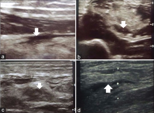
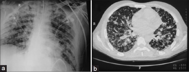
Pyomyositis is a tropical infection affecting skeletal muscles manifesting as high-grade fever with pain in the affected limbs usually caused by Gram-positive microorganisms. Gram-negative organisms causing pyomyositis is uncommon but has been reported. Burkholderia cepacia is a Gram-negative nonfermenter causing opportunistic infections in immunocompromised patients, has been reported to cause pyomyositis only once before. We report a case of B. cepacia pyomyositis in a patient with no history of immunocompromised status, manifesting as disseminated infection with hemophagocytic syndrome presenting to our intensive care unit.
Keywords: Burkholderia cepacia; disseminated infection; pyomyositis.
Conflict of interest statement
There are no conflicts of interest.
Figures

Similar articles
-
Acute Burkholderia cenocepacia pyomyositis in a patient with cystic fibrosis.J Cyst Fibros. 2009 Jul;8(4):273-5. doi: 10.1016/j.jcf.2009.04.007. Epub 2009 May 17. J Cyst Fibros. 2009. PMID: 19451037
-
Multidrug-resistant Burkholderia cepacia bacteremia in an immunocompetent adult diagnosed with dengue and scrub coinfection: A rare case report.Int J Crit Illn Inj Sci. 2018 Jul-Sep;8(3):173-175. doi: 10.4103/IJCIIS.IJCIIS_31_18. Int J Crit Illn Inj Sci. 2018. PMID: 30181976 Free PMC article.
-
[Burkholderia cepacia: a new pathogen causing nosocomial infections].Przegl Epidemiol. 2008;62(1):7-17. Przegl Epidemiol. 2008. PMID: 18536220 Polish.
-
Enterobacter cloacae pyomyositis complicating chronic granulomatous disease and review of gram-negative bacilli pyomyositis.Eur J Clin Microbiol Infect Dis. 2013 Jun;32(6):729-34. doi: 10.1007/s10096-013-1815-y. Epub 2013 Feb 1. Eur J Clin Microbiol Infect Dis. 2013. PMID: 23370968 Review.
-
Gram-negative bacterial pyomyositis: unique case and review.Rev Infect Dis. 1989 Sep-Oct;11(5):789-92. doi: 10.1093/clinids/11.5.789. Rev Infect Dis. 1989. PMID: 2682951 Review.
Cited by
-
Diabetic foot infection caused by bacteria of the Burkholderia cepacia complex: report of an unusual case and a scoping literature review.Rev Inst Med Trop Sao Paulo. 2022 Jun 6;64:e36. doi: 10.1590/S1678-9946202264036. eCollection 2022. Rev Inst Med Trop Sao Paulo. 2022. PMID: 35674634 Free PMC article.
References
-
- Chiedozi LC. Pyomyositis. Review of 205 cases in 112 patients. Am J Surg. 1979;137:255–9. - PubMed
-
- Walling DM, Kaelin WG., Jr Pyomyositis in patients with diabetes mellitus. Rev Infect Dis. 1991;13:797–802. - PubMed
-
- Gousseff M, Lanternier F, Ferroni A, Chandesris O, Mahlaoui N, Hermine O, et al. Enterobacter cloacae pyomyositis complicating chronic granulomatous disease and review of Gram-negative bacilli pyomyositis. Eur J Clin Microbiol Infect Dis. 2013;32:729–34. - PubMed
-
- El-Laboudi AH, Etherington C, Whitaker P, Clifton IJ, Conway SP, Denton M, et al. Acute Burkholderia cenocepacia pyomyositis in a patient with cystic fibrosis. J Cyst Fibros. 2009;8:273–5. - PubMed
-
- Liao CH, Chang HT, Lai CC, Huang YT, Hsu MS, Liu CY, et al. Clinical characteristics and outcomes of patients with Burkholderia cepacia bacteremia in an Intensive Care Unit. Diagn Microbiol Infect Dis. 2011;70:260–6. - PubMed
Publication types
LinkOut - more resources
Full Text Sources