. 2018 Dec 18:(808):123-160.
doi: 10.3897/zookeys.808.29679. eCollection 2018.

A taxonomic revision and molecular phylogeny of the eastern Palearctic species of the genera Schizomyia Kieffer and Asteralobia Kovalev (Diptera, Cecidomyiidae, Asphondyliini), with descriptions of five new species of Schizomyia from Japan

Affiliations

A taxonomic revision and molecular phylogeny of the eastern Palearctic species of the genera Schizomyia Kieffer and Asteralobia Kovalev (Diptera, Cecidomyiidae, Asphondyliini), with descriptions of five new species of Schizomyia from Japan

Ayman Khamis Elsayed et al. Zookeys. .

Abstract

The genus Asteralobia (Diptera, Cecidomyiidae, Asphondyliini, Schizomyiina) was erected by Kovalev (1964) based on the presence of constrictions on the cylindrical male flagellomeres. In the present study, we examine the morphological features of Asteralobia and Schizomyia and found that the male flagellomeres are constricted also in Schizomyiagaliorum, the type species of Schizomyia. Because no further characters clearly separating Asteralobia from Schizomyia were observed, we synonymize Asteralobia under Schizomyia. Molecular phylogenetic analysis strongly supports our taxonomic treatment. We describe five new species of Schizomyia from Japan, S.achyranthesae Elsayed & Tokuda, sp. n., S.diplocyclosae Elsayed & Tokuda, sp. n., S.castanopsisae Elsayed & Tokuda, sp. n., S.usubai Elsayed & Tokuda, sp. n., and S.paederiae Elsayed & Tokuda, sp. n., and redescribe three species, S.galiorum Kieffer, S.patriniae Shinji, and S.asteris Kovalev. A taxonomic key to the Japanese Schizomyia species is provided.

Keywords: Cecidomyiinae; Schizomyiina; gall midges; taxonomic key.


Figures

Figures 1–4.
Figures 1–4.
Galls of Schizomyia spp. 1 Fruit gall induced by S.achyranthesae on Achyranthesbidentata (Amaranthaceae) 2 Inflorescence galls induced by S.castanopsisae on Castanopsissieboldii (Fagaceae) 3 Fruit galls (arrows) induced by S.usubai on Trachelospermumasiaticum (Apocynaceae) 4 A flower bud gall (white arrow) induced by S.paederiae on Paederiafoetida (Rubiaceae) [red arrows indicate normal flower buds].
Figures 5–11.
Figures 5–11.
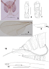
Schizomyiaachyranthesae. 5 Head 6 Ventral view of female antennal flagellomere V 7 Ventral view of male flagellomere V 8 Wing 9 Tarsomere V and acromere 10 Terminal part of female abdomen 11 Ovipositor apex. Scale bars: 50 µm (6, 7, 9–11), 200 µm (5, 8).
Figures 12–17.
Figures 12–17.
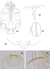
Schizomyiaachyranthesae. 12 Male terminalia 13 Larval spatula 14 Larval anus 15 Terminal larval segment dorsally 16 Ventral view of pupal head 17 Pupal prothoracic spiracle. Scale bars: 50 µm (12–15), 100 µm (16, 17).
Figures 18–24.
Figures 18–24.
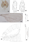
Schizomyiadiplocyclosae. 18 Head 19 Ventral view of female flagellomere V 20 Dorsal view of male flagellomere V 21 Wing 22 Tarsomere V and acromere 23 Terminal part of female abdomen 24 Ovipositor apex Scale bars: 100 µm (18, 19), 50 µm (20–24).
Figures 25–29.
Figures 25–29.
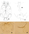
Schizomyiadiplocyclosae. 25 Male terminalia 26 Larval spatula 27 Terminal larval segments dorsally 28 Ventral view of pupal head 29 Prothoracic spiracle in pupa. Scale bars: 50 µm (25–27), 100 µm (28, 29).
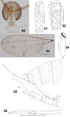
Figures 30–36.
Figures 30–36.
Schizomyiacastanopsisae. 30 Head 31 Ventral view of female flagellomere V 32 Ventral view of male flagellomere V 33 Wing 34 Tarsomere V and acromere 35 Terminal part of female abdomen 36 Ovipositor apex. Scale bars: 50 µm (31, 32, 34–36), 100 µm (30, 33).
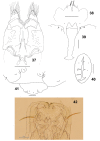
Figures 37–42.
Figures 37–42.
Schizomyiacastanopsisae. 37 Male terminalia 38 Male cerci 39 Larval spatula 40 Larval anus 41 Terminal larval segments dorsally 42 Ventral view of pupal head and thorax. Scale bars: 50 µm (37–41), 100 µm (42).
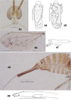
Figures 43–49.
Figures 43–49.
Schizomyiausubai. 43 Head 44 Ventral view of female flagellomere V 45 Ventral view of male flagellomere V 46 Wing 47 Tarsomere V and acromere 48 Terminal part of female abdomen 49 Ovipositor apex. Scale bars: 50 µm (44, 45, 47, 49), 100 µm (43, 46, 48).
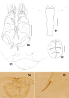
Figure 50–55.
Figure 50–55.
Schizomyiausubai. 50 Male terminalia 51 Larval spatula 52 Larval anus 53 Terminal larval segments dorsally 54 Ventral view of pupal head 55 Pupal prothoracic spiracle. Scale bars: 50 µm (50–53), 100 µm (54, 55).
Figures 56–62.
Figures 56–62.
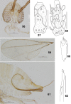
Schizomyiapaederiae. 56 Head 57 Dorsal view of female flagellomere V 58 Dorsal view of female flagellomere V 59 Wing 60 Tarsomere V and acromere 61 Terminal part of female abdomen 62 Ovipositor apex. Scale bars: 50 µm (57, 58, 60, 62), 100 µm (56, 59, 61).
Figures 63–67.
Figures 63–67.
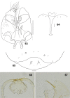
Schizomyiapaederiae. 63 Male terminalia 64 Larval spatula 65 Terminal larval segments dorsally 66 Ventral view of pupal head 67 Pupal prothoracic spiracle. Scale bars: 50 µm (63–65), 100 µm (66, 67).
Figures 68–73.
Figures 68–73.
Schizomyiagaliorum. 68 Lateral view of female flagellomere V 69 Lateral view of male flagellomere V 70 Wing 71 Tarsomere V and acromere 72 Terminal part of female abdomen showing the ovipositor 73 Ovipositor apex. Scale bars: 50 µm (68, 69, 71, 73), 100 µm (70, 72).
Figure 74–76.
Figure 74–76.
Schizomyiagaliorum. 74 Male terminalia 75 Ventral view of pupal head 76 Pupal prothoracic spiracle. Scale bars: 50 µm (74), 100 µm (75, 76).
Figures 77–83.
Figures 77–83.
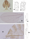
Schizomyiapatriniae. 77 Head 78 Dorsal view of female flagellomere V 79 Ventral view of female flagellomere V 80 Wing 81 Tarsomere V and acromere 82 Terminal part of female abdomen 83 Ovipositor apex. Scale bars: 50 µm (78, 79, 81, 83), 200 µm (77, 80, 82).
Figures 84–85.
Figures 84–85.
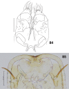
Schizomyiapaederiae. 84 Male terminalia 85 Ventral view of pupal head and prothorax. Scale bars: 50 µm (84), 200 µm (85).
Figures 86–93.
Figures 86–93.
Schizomyiaasteris. 86 Lateral view of female flagellomere V 87 Lateral view of male flagellomere V 88 Tarsomere V and acromere 89 Ovipositor apex 90 Male terminalia 91 Larval spatula 92 Terminal larval segments dorsally 93 Larval anus. Scale bar: 50 µm.
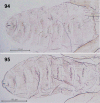
Figures 94–95.
Figures 94–95.
Dorsal view of pupal abdominal segments. 94Schizomyiasoyogo95Schizomyiasasaki. Scale bar: 100 µm.
Figure 96.
Figure 96.
A phylogenetic reconstruction based on partial sequences of cytochrome oxidase subunit I (COI) and 12S small ribosomal subunit genes. The topology and branch length were produced by the maximum likelihood method (note the scale bar). Bootstrap values are indicated at branches gaining more than 50% support (103 replications).

References

    1. Darriba D, Taboada GL, Doallo R, Posada D. (2012) jModelTest 2: more models, new heuristics and parallel computing. Nature Methods 9: 772. 10.1038/nmeth.2109 - DOI - PMC - PubMed
    1. Elsayed AK, Ogata K, Kaburagi K, Yukawa J, Tokuda M. (2017) A new Dasineura species (Diptera: Cecidomyiidae) associated with Symplocoscochinchinensis (Loureiro) (Symplocaceae) in Japan. Japanese Journal of Systematic Entomology 23: 81–86.
    1. Elsayed AK, Uechi N, Yukawa J, Tokuda M. (in press) Ampelomyia, a new genus of Schizomyiina (Diptera: Cecidomyiidae) associated with Vitis (Vitaceae) in the Palaearctic and Nearctic regions, with description of a new species from Japan. The Canadian Entomologist.
    1. Elsayed AK, Shimizu-Kaya U, Itioka T, Meleng P, Yukawa J, Tokuda M. (2018) A new genus and a new species of Schizomyiina (Diptera: Cecidomyiidae: Asphondyliini) inducing petiole galls on Macarangabancana (Miq.) in Borneo, Malaysia. Zootaxa 4482: 188–196. 10.11646/zootaxa.4482.1.10 - DOI - PubMed
    1. Fedotova ZA. (2002) New species of gall midges (Diptera, Cecidomyiidae) from the Russian Far East. Far Eastern Entomologist 118: 1–35.

LinkOut - more resources