Case Reports
. 2018 Nov 26:2018:4160925.
doi: 10.1155/2018/4160925. eCollection 2018.

Epiphyseal Primary Diffuse Large B-Cell Lymphoma of Bone

Affiliations
Case Reports

Epiphyseal Primary Diffuse Large B-Cell Lymphoma of Bone

Shachar Kenan et al. Case Rep Pathol. .

Abstract

Primary lymphoma of bone (PLB) confined to the epiphysis has only been described in four other patients. Due to the rarity of this entity, diagnosis has often been delayed, leading to mismanagement with adverse clinical consequences. We report a fifth case of primary epiphyseal lymphoma of bone located in the left distal medial femoral epiphysis of a 13-year-old boy. Radiographic and histologic features of PLB are discussed, along with a review of the literature and pitfalls of misdiagnosis. The patient initially presented with six months of progressive left knee pain with an associated loss of passive range of motion. Imaging revealed a mixed radiolucent lesion within the left distal medial femoral epiphysis with cortical breakthrough. A core biopsy was performed revealing a blue round cell tumor. Thanks to modern immunohistochemistry techniques, a diagnosis of primary lymphoma of bone was quickly made. The patient thus avoided further surgical intervention and received the appropriate treatment of chemotherapy, with subsequent rapid resolution of the lesion. This case highlights the necessity of including primary lymphoma of bone in all epiphyseal lesion differential diagnoses, especially in the pediatric patient population when aggressive radiographic features are present.


Figures

Figure 1
Figure 1
Computed tomography (CT): coronal (a), sagittal (b), and axial views (c) of the left knee, July 2009.
Figure 2
Figure 2
Magnetic resonance images (MRI): T1 coronal, sagittal, and axial views (a, b, c), STIR coronal (d), T2 sagittal (e), and axial views (f) of the left knee, July 2009.
Figure 3
Figure 3
Three-phase bone scintigraphy, August 2009. Increased uptake in the distal left femur on delayed images.
Figure 4
Figure 4
Fludeoxyglucose-positron emission tomography (FDG-PET), August 2009. Increased activity of FDG within the left knee (SUV max of 17.11), with physiologic distribution of FDG in the remainder of the body.
Figure 5
Figure 5
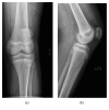
Anteroposterior (a) and lateral radiographs (b) of the left knee after core biopsy, September 2009.
Figure 6
Figure 6
High power magnification, hematoxylin and eosin (H&E) staining. Sheets of dyscohesive atypical cells with large hyperchromatic round nuclei and scant cytoplasm.
Figure 7
Figure 7
Immunohistochemistry staining. CD20 and CD79a: Strong positive cytoplasmic staining indicative of lymphoid cells of B-type lineage (a, b). CD3: scant scattered reactive T-cells in background (c).
Figure 8
Figure 8
Anteroposterior (a) and lateral (b) radiographs of the left knee after chemotherapy, October 2010. Dramatic lesion response after treatment.
Figure 9
Figure 9
Magnetic resonance images (MRI): T1 coronal, sagittal, axial views (a, b, c) and T1 fat saturated w/contrast coronal, sagittal, axial views (d, e, f) of the left knee after completion of chemotherapy, October 2010. Near complete resolution of lesion.
Figure 10
Figure 10
Fludeoxyglucose-positron emission tomography (FDG-PET), May 2011. Non-FDG-avid mixed sclerotic and lytic lesion of the left medial femoral condyle, consistent with resolved lymphoma.

References

    1. Jawad M. U., Schneiderbauer M. M., Min E. S., Cheung M. C., Koniaris L. G., Scully S. P. Primary lymphoma of bone in adult patients. Cancer. 2010;116(4):871–879. doi: 10.1002/cncr.24828. - DOI - PubMed
    1. Cai L., Stauder M. C., Zhang Y.-J., et al. Early-stage primary bone lymphoma: a retrospective, multicenter Rare Cancer Network (RCN) study. International Journal of Radiation Oncology, Biology, Physics. 2012;83(1):284–291. doi: 10.1016/j.ijrobp.2011.06.1976. - DOI - PubMed
    1. Kitsoulis P., Vlychou M., Papoudou-Bai A., et al. Primary lymphomas of bone. Anticancer Reseach. 2006;26(1):325–337. - PubMed
    1. Hogendoorn P. C. W., Kluin P. M. Primary non-Hodgkin lymphoma of bone. In: Fletcher C. D. M., Bridge J. A., Hogendoorn P. C. W., Mertens F., editors. WHO Classification of Tumours of Soft Tissue and Bone. 4th 2013.
    1. Thawait S. K., Thawait G. K., Frassica F. J., Andreisek G., Carrino J. A., Chhabra A. A systematic approach to magnetic resonance imaging evaluation of epiphyseal lesions. Magnetic Resonance Imaging. 2013;31(3):418–431. doi: 10.1016/j.mri.2012.08.006. - DOI - PubMed

Publication types

LinkOut - more resources