

Pediatric Transthoracic Cardiac Vector Flow Imaging - A Preliminary Pictorial Study
- PMID: 30599042
- PMCID: PMC6303157
- DOI: 10.1055/a-0656-5430
Pediatric Transthoracic Cardiac Vector Flow Imaging - A Preliminary Pictorial Study
Abstract
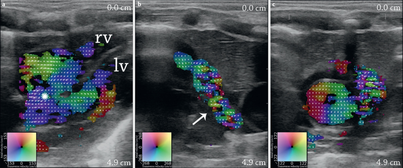
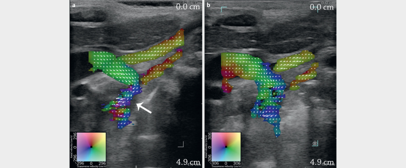
Purpose Conventional pediatric echocardiography is crucial for diagnosing congenital heart disease (CHD), but the technique is impaired by angle dependency. Vector flow imaging (VFI) is an angle-independent noninvasive ultrasound alternative for blood flow assessment and can assess complex flow patterns not visible on conventional Doppler ultrasound. Materials and Methods 12 healthy newborns and 3 infants with CHD were examined with transthoracic cardiac VFI using a conventional ultrasound scanner and a linear array. Results VFI examinations revealed common cardiac flow patterns among the healthy newborns, and flow changes among the infants with CHD not previously reported with conventional echocardiography. Conclusion For assessment of cardiac flow in the normal and diseased pediatric heart, VFI may provide additional information compared to conventional echocardiography and become a useful diagnostic tool.
Keywords: Cardiac flow; Congenital heart disease; Transthoracic echocardiography; Transverse Oscillation; Vector Flow Imaging.
Conflict of interest statement
Figures


References
-
- van der Linde D, Konings E E, Slager M A et al. Birth prevalence of congenital heart disease worldwide: A systematic review and meta-analysis. J Am Coll Cardiol. 2011;58:2241–2247. - PubMed
-
- Bharucha T, Mertens L. Recent advances in pediatric echocardiography. Expert Rev Cardiovasc Ther. 2013;11:31–47. - PubMed
-
- Deeg K H. Echocardiographic differential diagnosis of the cyanotic newborn. Ultraschall in Med. 2015;36:104–118. - PubMed
-
- Berg A, Greve G. Trends in pediatric imaging: Ultrasound. Acta Radiol. 2013;54:1096–1105. - PubMed
-
- Evans D H, McDicken N, Skidmore R . New York: John Wiley & Sons; 1989. Doppler ultrasound, Physics.Instrumentation and Clinical Applications.
LinkOut - more resources
Full Text Sources