. 2019 Jan;37(1):139-149.
doi: 10.1002/stem.2921.

The Role of Nerve Growth Factor in Maintaining Proliferative Capacity, Colony-Forming Efficiency, and the Limbal Stem Cell Phenotype

Affiliations

The Role of Nerve Growth Factor in Maintaining Proliferative Capacity, Colony-Forming Efficiency, and the Limbal Stem Cell Phenotype

Sai Kolli et al. Stem Cells. 2019 Jan.

Abstract

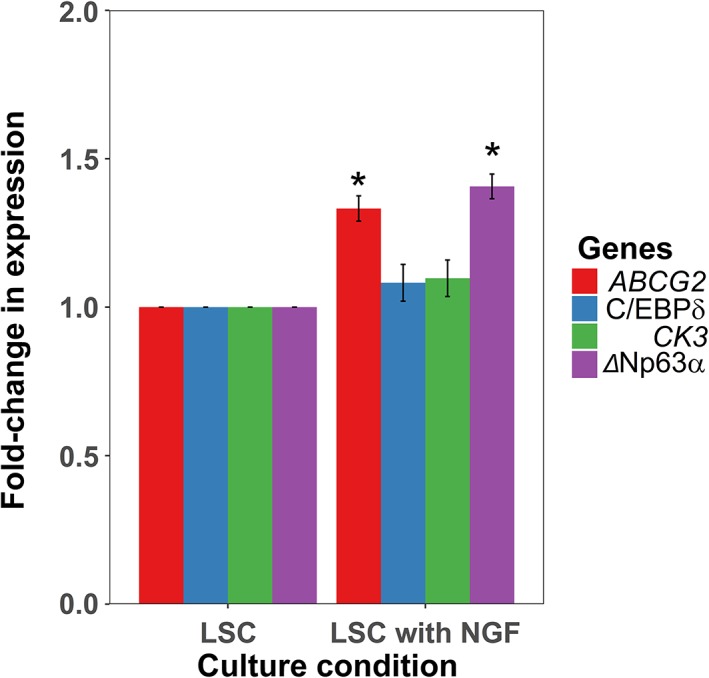
Nerve growth factor (NGF) has demonstrated great benefit in the treatment of neurotrophic corneal ulcers. There is evidence for multiple modes of action in promoting corneal healing, but only indirect evidence exists for NGF's effects on limbal stem cells (LSCs). Understanding the role of NGF in LSC biology will improve our understanding of paracrine regulation of the limbal niche and the design of stem cell-based therapies for conditions such as LSC deficiency. In this article, we studied the regulation of NGF signaling components during LSC differentiation and the role of NGF in LSC proliferation and maintenance of the stem cell phenotype. LSC differentiation was induced by prolonged (40 day) culture which resulted in a significant increase in cell size, decrease in colony-forming efficiency and expression of putative LSC markers. A protein microarray measuring expression of 248 signaling proteins indicated the low affinity NGF receptor p75NTR to be the most downregulated protein upon differentiation. Further confirmation by Western blotting and real-time quantitative polymerase chain reaction indicated that NGF and p75NTR are expressed in early LSC cultures and downregulated upon differentiation. LSC cultures grown in the presence of anti-NGF antibody showed decreased colony-forming efficiency, DNA replication and expression of putative LSC markers ABCG2 and C/EBPδ. Supplementation of LSC culture medium with NGF extended the life span of LSC cultures in vitro and increased the expression of putative LSC markers ΔNp63α and ABCG2. Taken together, our data indicate that NGF signaling is a key promoter of LSC proliferation, colony-forming efficiency, and a maintainer of the LSC phenotype. Stem Cells 2019;37:139-149.

Keywords: Colony-forming efficiency; Corneal epithelium; Limbal stem cell deficiency; Limbal stem cell markers; Limbal stem cells; Nerve growth factor; Proliferation; Stem cell niche.


Figures

Figure 1
Figure 1
Schematic representation of corneal epithelial stem cell differentiation protocol and sample collection time points. Abbreviations: RT‐qPCR, real‐time quantitative polymerase chain reaction; CFE, colony‐forming efficiency.
Figure 2
Figure 2
Limbal stem cells (LSC) differentiation results in reduced colony‐forming efficiency (CFE), reduced expression of putative LSC markers, and increased expression of putative markers of differentiated corneal epithelium. (A): Bar chart showing the results of the CFE assay during differentiation protocol. There is a significant reduction in CFE during the differentiation protocol (p < .001; n = 3; one‐way analysis of variance (ANOVA); SEM shown by error bars). (B): Real‐time quantitative polymerase chain reaction (RT‐qPCR) of putative LSC markers and a corneal differentiation marker at different stages after primary culture. There is a statistically significant decrease in putative LSC markers ABCG2, C/EBPδ, ΔNp63α (p < .000001, <.001, and < .000001, respectively; n = 3; one‐way ANOVA; SEM) and an increase in differentiation marker CK3 throughout the culture process (p < .001; n = 3; one‐way ANOVA; SEM). Between‐group analyses are shown in Supporting Information Table S4. Abbreviation: GAPDH, glyceral‐dehyde‐3‐phosphate dehydrogenase.
Figure 3
Figure 3
Bioinformatics analysis of differentially expressed proteins between the stem cell and differentiated states shows that cell cycle regulation and apoptosis are enriched biological process terms. NGF signalling is a highly enriched pathway term and potentially controls several other pathways known to be involved in corneal healing. (A) Top 20 ranked biological functions by combined p‐value and Z‐score (Enrichr) against corresponding negative log p‐values. The orange line represents a p‐value = 0.05. A list of genes coding for the proteins analysed in the Panorama Cell Signalling Protein microarray was drawn up. Genes which met the 1.2‐fold change cut‐off were used in the analysis of gene ontology. (B) Dot‐plot representing the significance of the top 20 enriched pathways ranked by number of genes involved (ReactomePA). Genes are clustered into pathway terms based on membership of Reactome pathways. Count is the number of genes involved in the pathway. Gene Ratio is the ratio of the genes involved in the pathway to the total number of genes analysed. P adjust is the adjusted p‐value. (C) Functional enrichment map of the top 20 enriched pathways by number of genes involved (ReactomePA). Nodes represent Reactome pathway terms. Edges between pathway terms represent overlap of pathway members.
Figure 4
Figure 4
Expression of nerve growth factor (NGF) and its receptors during the differentiation of limbal stem cells. (A): Western blot analysis of TrkA, p75NTR, NGF, with β‐actin (43 kDa) as a housekeeping control at days 10, 20, 30, 40, and 50 of culture. Mature NGF was detected as two bands at 27 and 13 kDa. Two bands likely representing two glycosylation forms of p75NTR at 62 and 83 kDa were also found as well as a band at 145 kDa representing TrkA. p75NTR and NGF decreased with differentiation while TrkA expression was variable with no clear trend. Full blots are shown in Supporting Information Figure S2A. (B): RT‐qPCR of NGF, p75NTR, and TrkA at days 10, 20, 30, and 40 of culture showing a significant decrease of NGF and p75NTR during differentiation (p < 1 × 10⁻⁹ and p < 1 × 10⁻⁵, respectively).
Figure 5
Figure 5
Nerve growth factor (NGF) blocking of limbal epithelial cultures decreases putative limbal stem cell (LSC) markers, colony‐forming efficiency (CFE), and proliferation. (A): Schema showing experimental set up for anti‐NGF studies. Each well shown was set up in biological triplicate. (B): Microphotographs of LSC culture at day 10 of culture in standard conditions (i) and in the presence of anti‐NGF (ii). Under a light microscope, the culture appearances are similar with tightly packed cells that are regular with large nuclei and scant cytoplasm. (C): There is no significant difference in mean cell area, as measured at day 10 of culture when LSCs are cultured in the presence of anti‐NGF (p = .43; n = 3; Student's t test; SEM). (D): CFE assay of standard culture versus standard culture with anti‐NGF at day 14 of culture. Culture in the presence of anti‐NGF results in a statistically significant reduction in CFE (p = .039; n = 3; Student's t test; SEM). (E): Bar charts showing RT‐qPCR results for the putative LSC markers ABCG2, C/EBPδ, ΔNp63α, and the corneal differentiation marker CK3 at day 14 of culture. There is a statistically significant decrease in putative LSC markers ABCG2 and C/EBPδ at day 14 of culture (p = .03 and .04, respectively; n = 3; Student's t test; SEM). There is a downward trend of ΔNp63α expression, and an upward trend of differentiation marker CK3 when LSCs are cultured in the presence of anti‐NGF (p = .15 and .53, respectively; n = 3; Student's t test; SEM). (F): There is a statistically significant decrease in proliferation of LSCs cultured in the presence of anti‐NGF as measured at day 14 of culture (p = .042; n = 3; Student's t test; SEM). (G): There is no significant change in the levels of apoptosis when cells are cultured in the presence of anti‐NGF, as measured by APO‐DIRECT flow cytometry at day 14 of culture. There are very few apoptotic cells in both culture conditions, showing that the concentration of NGF was not causing direct toxicity to the cells (p = .12; n = 3; Student's t test; SEM).
Figure 6
Figure 6
Nerve growth factor (NGF) addition to limbal epithelial cultures increases putative limbal stem cell (LSC) markers. Bar charts showing RT‐qPCR results for the putative LSC markers ABCG2, C/EBPδ, ΔNp63α, and the corneal differentiation marker CK3 at day 40 of culture. There is a statistically significant increase in putative LSC markers ΔNp63α and ABCG2 (p < 1 × 10⁻⁷ and p < 1 × 10⁻⁶, respectively; n = 3; Student's t test; SEM) whereas CEPBδ and CK3 levels do not show significant differences between the groups (p = .099 and .104, respectively; n = 3; Student's t test; SEM).

References

    1. Osei‐Bempong C, Figueiredo FC, Lako M. The limbal epithelium of the eye—A review of limbal stem cell biology, disease and treatment. Bioessays 2013;35:211–219. - PubMed
    1. Ebato B, Friend J, Thoft RA. Comparison of limbal and peripheral human corneal epithelium in tissue culture. Investig Ophthalmol Vis Sci 1988;29:1533–1537. - PubMed
    1. Mann I. A study of epithelial regeneration in the living eye. Br J Ophthalmol 1944;28:26–40. - PMC - PubMed
    1. Mann I. A study of eighty‐four cases of delayed mustard gas keratitis fitted with contact lenses. Br J Ophthalmol 1944;28:441–447. - PMC - PubMed
    1. Davanger M, Evensen A. Role of the pericorneal papillary structure in renewal of corneal epithelium. Nature 1971;229:560–561. - PubMed

Publication types

Substances