. 2019 Mar;290(3):702-708.
doi: 10.1148/radiol.2018180556. Epub 2019 Jan 1.

Compressed Sensing Radial Sampling MRI of Prostate Perfusion: Utility for Detection of Prostate Cancer

Affiliations

Compressed Sensing Radial Sampling MRI of Prostate Perfusion: Utility for Detection of Prostate Cancer

David J Winkel et al. Radiology. 2019 Mar.

Abstract

Purpose To investigate the diagnostic performance of a dual-parameter approach by combining either volumetric interpolated breath-hold examination (VIBE)- or golden-angle radial sparse parallel (GRASP)-derived dynamic contrast agent-enhanced (DCE) MRI with established diffusion-weighted imaging (DWI) compared with traditional single-parameter evaluations on the basis of DWI alone. Materials and Methods Ninety-four male participants (66 years ± 7 [standard deviation]) were prospectively evaluated at 3.0-T MRI for clinical suspicion of prostate cancer. Included were 101 peripheral zone prostate cancer lesions. Histopathologic confirmation at MRI transrectal US fusion biopsy was matched with normal contralateral prostate parenchyma. MRI was performed with diffusion weighting and DCE by using GRASP (temporal resolution, 2.5 seconds) or VIBE (temporal resolution, 10 seconds). Perfusion (influx forward volume transfer constant [Ktrans] and rate constant [Kep]) and apparent diffusion coefficient (ADC) parameters were determined by tumor volume analysis. Areas under the receiver operating characteristic curve were compared for both sequences. Results Evaluated were 101 prostate cancer lesions (GRASP, 61 lesions; VIBE, 40 lesions). In a combined analysis, diffusion and perfusion parameters ADC with Ktrans or Kep acquired with GRASP had higher diagnostic performance compared with diffusion characteristics alone (area under the curve, 0.97 ± 0.02 [standard error] vs 0.93 ± 0.03; P < .006 and .021, respectively), whereas ADC with perfusion parameters acquired with VIBE had no additional benefit (area under the curve, 0.94 ± 0.03 vs 0.93 ± 0.04; P = .18and .50, respectively, for combination of ADC with Ktrans and Kep). Conclusion If used in a dual-parameter model, incorporating diffusion and perfusion characteristics, the golden-angle radial sparse parallel acquisition technique improves the diagnostic performance of multiparametric MRI examinations of the prostate. This effect could not be observed combining diffusing with perfusion parameters acquired with volumetric interpolated breath-hold examination. © RSNA, 2018.


Conflict of interest statement

Disclosures of Conflicts of Interest:

D.J.W. disclosed no relevant relationships. T.J.H. disclosed no relevant relationships. M.R.B. disclosed no relevant relationships. C.G.G. disclosed no relevant relationships. C.W. disclosed no relevant relationships. L.B. disclosed no relevant relationships. T.K.B. Activities related to the present article: disclosed no relevant relationships. Activities not related to the present article: disclosed money to author’s institution for patent issued for GRASP technique. Other relationships: disclosed no relevant relationships. D.T.B. disclosed no relevant relationships.

Figures

Figure 1:
Figure 1:
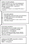
Flowchart outlines the selection of the final study population with inclusion and exclusion criteria within the defined observation window. DCE = dynamic contrast-agent enhanced, GRASP = golden-angle radial sparse parallel, mpMRI = multiparametric MRI, PCa = prostate cancer, PSA = prostate-specific antigen, VIBE = volumetric interpolated breath-hold examination.
Figure 2:
Figure 2:
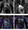
Perfusion data set acquired at volumetric interpolated breath-hold examination. Images in a 68-year-old man with a Gleason score of 8 (4 + 4) and prostate-specific antigen level of 7.4 (mg/L). (a) T2-weighted MR image shows hypointense signal of the prostate cancer lesion. (b) Apparent diffusion coefficient (ADC) map shows hypointense signal of the prostate cancer lesion with an ADC value of 0.91 mm2/sec ± 0.29. (c) Perfusion map shows influx forward volume transfer constant (Ktrans) value of 0.26 min21 ± 0.15. (d) Perfusion map shows rate constant (Kep) value of 0.46 min−1 ± 0.30. Volume of interest = 2.13 cm3. Note the extension of volume-of-interest boundaries (green outlines) defined on ADC series into interface regions between normal and pathologic parenchyma on dynamic contrast-enhanced MR images.
Figure 3:
Figure 3:
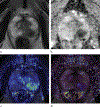
Perfusion data set acquired by using golden-angle radial sparse parallel method. Images in a 67-year-old man with a Gleason score of 8 (4 + 4) and a prostate-specific antigen level of 18.15 mg/L. (a) T2-weighted MR image shows hypointense signal of the prostate cancer lesion. (b) Apparent diffusion coefficient (ADC) map shows hypointense signal in the prostate cancer lesion; ADC = 1.12 mm2/sec ± 0.39. (c) Perfusion map shows influx forward volume transfer constant (Ktrans; 0.25 min−1 ± 0.21). (d) Perfusion map shows rate constant (Kep; 1.79 min−1 ± 0.97). Lesion volume = 2.32 cm3. There is an extension of volume-of-interest boundaries (green outlines) defined on ADC series into interface regions between normal and pathologic parenchyma on dynamic contrast-enhanced MR images.
Figure 4:
Figure 4:
Quantitative assessment of combined diffusion MRI and dynamic contrast agent–enhanced MRI. Scatterplots display the data pairs of apparent diffusion coefficients (ADCs) with (a) influx forward volume transfer constant and (b) ADC with rate constant values. ADC values are on the x-axis and the perfusion parameters are on the y-axis. Horizontal and vertical lines represent the dual-parameter cutoff levels. Note that the cutoff levels on the basis of golden-angle radial sparse parallel (GRASP)–derived perfusion parameters combined with ADC allows a better differentiation (ie, less overlap) between normal and prostate parenchyma compared to cutoff levels on the basis of volumetric interpolated breath-hold examination (VIBE)–derived perfusion parameters combined with ADC.

References

    1. Lüdemann L, Prochnow D, Rohlfing T, et al. Simultaneous quantification of perfusion and permeability in the prostate using dynamic contrast-enhanced magnetic resonance imaging with an inversion-prepared dual-contrast sequence. Ann Biomed Eng 2009;37(4):749–762. - PubMed
    1. van Niekerk CG, van der Laak JAWM, Hambrock T, et al. Correlation between dynamic contrast-enhanced MRI and quantitative histopathologic microvascular parameters in organ-confined prostate cancer. Eur Radiol 2014;24(10):2597–2605. - PubMed
    1. Langer DL, van der Kwast TH, Evans AJ, Trachtenberg J, Wilson BC, Haider MA. Prostate cancer detection with multi-parametric MRI: Logistic regression analysis of quantitative T2, diffusion-weighted imaging, and dynamic contrast-enhanced MRI. J Magn Reson Imaging 2009;30(2):327–334. - PubMed
    1. Giannini V, Mazzetti S, Armando E, et al. Multiparametric magnetic resonance imaging of the prostate with computer-aided detection: experienced observer performance study. Eur Radiol 2017;27(10):4200–4208. - PubMed
    1. Rosenkrantz AB, Geppert C, Grimm R, et al. Dynamic contrast-enhanced MRI of the prostate with high spatiotemporal resolution using compressed sensing, parallel imaging, and continuous golden-angle radial sampling: preliminary experience. J Magn Reson Imaging 2015;41(5):1365–1373. - PMC - PubMed