. 2018 Dec;15(4):394-399.
doi: 10.14245/ns.1836056.028. Epub 2018 Oct 2.

The Effectiveness of Noninvasive Positive Pressure Ventilation in Subarachnoid Pleural Fistula: A Case Report and Literature Review

Affiliations

The Effectiveness of Noninvasive Positive Pressure Ventilation in Subarachnoid Pleural Fistula: A Case Report and Literature Review

Dilip Chand Raja Soundararajan et al. Neurospine. 2018 Dec.

Abstract

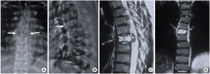
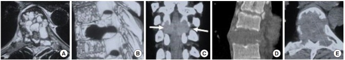
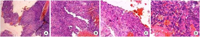
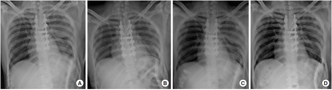
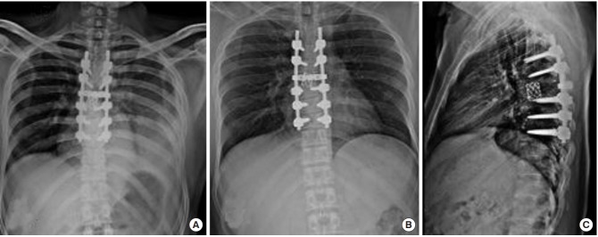
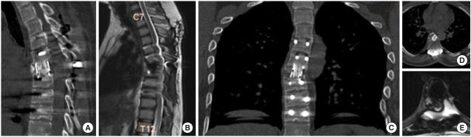
Subarachnoid pleural fistula (SPF) is an aberrant communication between the pleural cavity and subarachnoid space, resulting in uncontrolled cerebrospinal fluid drainage. The negative pressure of the pleural cavity creates a continuous suctioning effect, thereby impeding the spontaneous closure of these fistulas. Dural tears or punctures in cardiothoracic procedures, spinal operations, and trauma are known to cause such abnormal communications. Failure to recognize this entity may result in sudden neurological or respiratory complications. Hence, a high index of suspicion is required for early diagnosis and prompt management. Noninvasive positive pressure ventilation has been described to be effective in managing such fistulas, thus mitigating the high morbidity associated with exploratory surgery for primary repair. Herein, we describe the typical presentation of SPF and the clinical course, treatment, and follow-up of a patient who sustained SPF following anterior thoracic spinal surgery.

Keywords: Incidental dural tear; Noninvasive positive pressure ventilation; Subarachnoid pleural fistula.


Conflict of interest statement

The authors have nothing to disclose.

Figures

Fig. 1.
Fig. 1.
(A) Anteroposterior radiograph: arrows point toward loss of pedicle margins on both sides and osteolytic lesion involving the entire D6 vertebra. (B) Lateral radiograph shows mild collapse of the vertebra. (C, D) Sagittal and coronal images of T2-weighted magnetic resonance imaging show an expansile lytic lesion with thinned out sclerotic margin and multiple fluid levels replacing the D6 vertebral body, along with retropulsion and soft tissue expansion of lesion posteriorly into the spinal canal resulting in secondary spinal canal stenosis and cord edema.
Fig. 2.
Fig. 2.
(A) Axial T2-weighted magnetic resonance imaging image shows multiple fluid pockets involving the vertebral body, adjacent pedicles and left lamina with posterior extension of lesion causing spinal canal compromise. (B) Sagittal 3-dimensional computed tomography (CT) image shows cortical breech, with posterior and epidural extension of the lesion. (C) Coronal CT arrows marks show distortion of pedicle margins bilaterally. (D) Sagittal CT image showing lytic lesion involving the entire height of vertebral body. (E) Axial CT cut shows thinned out sclerotic margins in anterior aspect of the lesion and grossly thinned out margins in the rest of vertebra.
Fig. 3.
Fig. 3.
Histopathology slides (H&E, × 100). (A) Cystic spaces of varying sizes are filled with blood lined by plump spindle-shaped mononuclear cells and they are separated by cellular stroma. (B) Cystic vascular spaces are lined by fibroblasts and are devoid of endothelium. (C) Vascular space is lined by fibroblasts and the septa shows few osteoclastic giant cells without any evidence of atypia/necrosis/increased mitosis or granulomata. (D) Clusters of multinucleated osteoclastic giant cells seen admixed with spindle stromal cells. The giant cells are small with 3 to 6 nuclei and are lined along the septa. The above images are suggestive of aneurysmal bone cyst.
Fig. 4.
Fig. 4.
Serial chest radiographs with intercostal chest drain (ICD) in situ demonstrating resolution of pleural effusion. (A) Massive pleural effusion on fourth postoperative day (POD). (B) Second day after initiating noninvasive positive pressure ventilation (NPPV) shows reappearance of basal lung air shadows. (C) Lung margins are appreciated better in this image taken 2 days later. (D) Complete resolution of haziness on day 5 of NPPV. ICD was removed on POD 9.
Fig. 5.
Fig. 5.
(A) Reappearance of minimal basal pleural effusion after removing intercostal chest drain (ICD) on day 6 after noninvasive positive pressure ventilation. (B, C) Complete resolution of pleural effusion with no recurrence after three months with implant cage in good position.
Fig. 6.
Fig. 6.
(A, B) Sagittal computed tomography (CT) image shows good fusion and bone to bone healing after 6 months with no evidence of recurrence. (C) Coronal magnetic resonance imaging (MRI) image shows clear chest fields with no recurrence of subarachnoid pleural fistula. Axial CT (D) and MRI (E) images show mesh cage in position and adequate decompression of cord.

References

    1. Fountas KN, Kapsalaki EZ, Johnston KW. Cerebrospinal fluid fistula secondary to dural tear in anterior cervical discectomy and fusion: case report. Spine (Phila Pa 1976) 2005;30:E277–80. - PubMed
    1. Milloy FJ, Correll NO, Langston HT. Persistent subarachnoid-pleural space fistula; report of a case. J Am Med Assoc. 1959;169:1467. - PubMed
    1. D'Souza R, Doshi A, Bhojraj S, et al. Massive pleural effusion as the presenting feature of a subarachnoid-pleural fistula. Respiration. 2002;69:96–9. - PubMed
    1. Khurana A, Brousil J, Russo A, et al. Intracranial hypotension with a sixth cranial nerve palsy subsequent to massive thoracic CSF hygroma: a rare complication of thoracic disc excision. Eur Spine J. 2013;22:2047–54. - PMC - PubMed
    1. Assietti R, Kibble MB, Bakay RA. Iatrogenic cerebrospinal fluid fistula to the pleural cavity: case report and literature review. Neurosurgery. 1993;33:1104–8. - PubMed

LinkOut - more resources