

Gadolinium-enhanced MRI reveals dynamic development of endolymphatic hydrops in Ménière's disease
- PMID: 30600169
- PMCID: PMC9422425
- DOI: 10.1016/j.bjorl.2018.10.014
Gadolinium-enhanced MRI reveals dynamic development of endolymphatic hydrops in Ménière's disease
Abstract
Introduction: Meniere's disease is associated with impaired hearing, tinnitus, vertigo, and aural fullness. Many anatomical studies have suggested idiopathic endolymphatic hydrops as the pathological basis of Meniere's disease, which now can be visualized by using gadolinium -enhanced magnetic resonance imaging of the inner ear.
Objective: To investigate the development of endolymphatic hydrops in Meniere's disease by monitoring the vestibules and cochleae of affected patients.
Methods: Inner ears of 178 patients with definite unilateral Meniere's disease diagnosis were visualized by 3-dimensional fluid-attenuated inversion recovery and three-dimensional real inversion recovery magnetic resonance imaging following bilateral gadolinium intratympanic injection. The scans were used to evaluate the presence and degree of endolymphatic hydrops in the vestibules and cochlear structures, including the cochlear apical turn, the cochlear middle turn, and the cochlear basal turn. The correlation of endolymphatic hydrops occurrence between the various parts of the inner ear was determined.
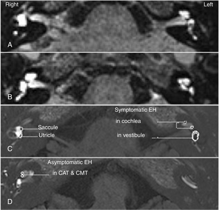
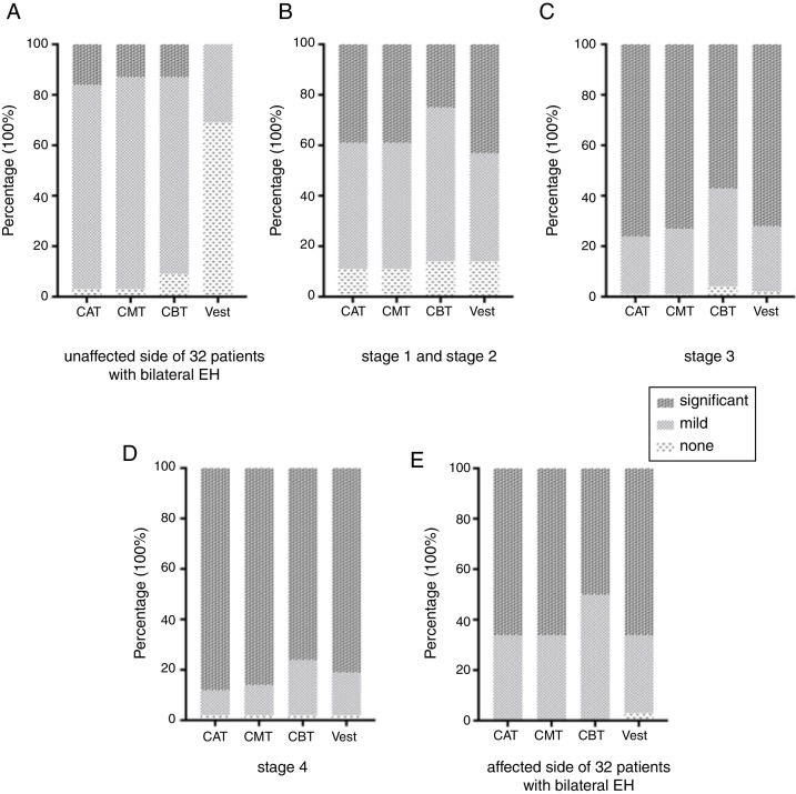
Results: Symptomatic endolymphatic hydrops was detected on the affected side in all patients, whereas asymptomatic endolymphatic hydrops was detected on the unaffected contra-lateral side in 32 patients (18.0%). On the affected side, the cochlear apical turn and the cochlear middle turn demonstrated significantly higher rates of endolymphatic hydrops than the cochlear basal turn and the vestibule. The severity of endolymphatic hydrops gradually decreased from the cochlear apical turn to the cochlear basal turn. On the contra lateral side, the incidence and degree of the detected asymptomatic endolymphatic hydrops were significantly greater in the cochleae than in the vestibules (p<0.05), with no significant difference detected between the cochlear turns.
Conclusion: Progression of endolymphatic hydrops appears to be directional, initiated in the cochlea. The order of endolymphatic hydrops severity gradually decreases from the cochlear apical turn to the cochlear basal turn and then to the vestibule. Endolymphatic hydrops in the vestibule is associated with symptomatic Meniere's disease.
Introdução: A doença de Ménière está associada a deficiência auditiva, zumbido, vertigem e plenitude auricular. Muitos estudos anatômicos sugerem hidropsia endolinfática idiopática como a base patológica da doença, que agora pode ser visualizada através de estudo por imagem da orelha interna por ressonância magnética com gadolínio.
Objetivo: Investigar o desenvolvimento da hidropsia endolinfática na doença de Ménière com monitoramento dos vestíbulos e das cócleas dos pacientes afetados.
Métodos: Orelhas internas de 178 pacientes com diagnóstico definitivo de doença de Ménière unilateral foram visualizados através de imagem de recuperação de inversão atenuada por fluidos em ressonância magnética tridimensional, 3-D FLAIR, e por inversão real após injeção intratimpânica bilateral de gadolínio. Os exames foram usados para avaliar a presença e o grau de hidropsia endolinfática nos vestíbulos e nas estruturas cocleares, inclusive o giro coclear apical, o giro coclear médio e o giro coclear basal. A correlação da ocorrência de hidropsia endolinfática entre as várias partes da orelha interna foi determinada.
Resultados: Hidropsia endolinfática sintomática foi detectada no lado afetado em todos os pacientes, enquanto hidropsia endolinfática assintomática foi detectada no lado contralateral não afetado em 32 pacientes (18,0%). No lado afetado, o giro apical da cóclea e o giro coclear médio demonstraram taxas significativamente mais altas de hidropsia endolinfática do que o giro basal e o vestíbulo. A gravidade da hidropsia endolinfática diminuiu gradualmente do giro apical da cóclea para o giro basal. No lado contralateral, a incidência e o grau da hidropsia endolinfática assintomática detectada foram significantemente maiores nas cócleas do que nos vestíbulos (p < 0,05), sem diferença significante entre os giros cocleares.
Conclusões: A progressão da hidropsia endolinfática parece ser direcional, iniciando-se na cóclea. A sua ordem da gravidade diminui gradualmente do giro apical da cóclea para o giro basal e, em seguida, para o vestíbulo. A hidropsia endolinfática no vestíbulo está associada à doença de Ménière sintomática.
Keywords: Doença de Ménière; Endolymphatic hydrops; Gadolinium; Gadolínio; Hidropsia endolinfática; Imagem de ressonância magnética; Injection; Injeção; Intratimpânica; Intratympanic; Magnetic resonance imaging; Meniere disease.
Copyright © 2018 Associação Brasileira de Otorrinolaringologia e Cirurgia Cérvico-Facial. Published by Elsevier Editora Ltda. All rights reserved.
Figures
Similar articles
-
[Visualization of endolymphatic hydrops in 3D-FLAIR MRI after intratympanic Gd-DTPA administration in Meniere's disease patients].Zhonghua Er Bi Yan Hou Tou Jing Wai Ke Za Zhi. 2013 Aug;48(8):628-33. Zhonghua Er Bi Yan Hou Tou Jing Wai Ke Za Zhi. 2013. PMID: 24195817 Chinese.
-
The Correlation Between Endolymphatic Hydrops and blood-labyrinth barrier Permeability of Meniere Disease.Ann Otol Rhinol Laryngol. 2021 Jun;130(6):578-584. doi: 10.1177/0003489420964823. Epub 2020 Oct 13. Ann Otol Rhinol Laryngol. 2021. PMID: 33047609
-
Diagnosis of endolymphatic hydrops by means of 3T magnetic resonance imaging after intratympanic administration of gadolinium.Radiologia. 2017 Mar-Apr;59(2):159-165. doi: 10.1016/j.rx.2016.10.006. Epub 2016 Dec 23. Radiologia. 2017. PMID: 28017456 English, Spanish.
-
MR imaging of endolymphatic hydrops in Ménière's disease: not all that glitters is gold.Acta Otorhinolaryngol Ital. 2018 Aug;38(4):369-376. doi: 10.14639/0392-100X-1986. Acta Otorhinolaryngol Ital. 2018. PMID: 30197428 Free PMC article. Review.
-
Magnetic resonance imaging and Ménière's disease-unavoidable alliance.Neuroradiology. 2021 Nov;63(11):1749-1763. doi: 10.1007/s00234-021-02744-5. Epub 2021 Jun 17. Neuroradiology. 2021. PMID: 34142211 Review.
Cited by
-
The diagnostic performance of cochlear endolymphatic hydrops and perilymphatic enhancement in stratifying Ménière's disease probabilities: A meta-analysis of semi-quantitative MRI-based grading systems.PLoS One. 2024 Nov 21;19(11):e0310045. doi: 10.1371/journal.pone.0310045. eCollection 2024. PLoS One. 2024. PMID: 39570877 Free PMC article.
-
The Third Mobile Window Syndrome: A Clinical Spectrum of Different Anatomical Locations-Characterization, Therapeutic Response, and Implications in the Development of Endolymphatic Hydrops.J Clin Med. 2024 Nov 28;13(23):7232. doi: 10.3390/jcm13237232. J Clin Med. 2024. PMID: 39685691 Free PMC article.
-
Delayed post gadolinium MRI descriptors for Meniere's disease: a systematic review and meta-analysis.Eur Radiol. 2023 Oct;33(10):7113-7135. doi: 10.1007/s00330-023-09651-8. Epub 2023 May 12. Eur Radiol. 2023. PMID: 37171493 Free PMC article.
-
Value of Endolymphatic Hydrops and Perilymph Signal Intensity in Suspected Ménière Disease.AJNR Am J Neuroradiol. 2020 Mar;41(3):529-534. doi: 10.3174/ajnr.A6410. Epub 2020 Feb 6. AJNR Am J Neuroradiol. 2020. PMID: 32029469 Free PMC article.
-
Endolymphatic hydrops imaging and correlation with clinical characteristics, audiovestibular function and mental impairment in patients with Meniere's disease.Eur Arch Otorhinolaryngol. 2023 Sep;280(9):4027-4036. doi: 10.1007/s00405-023-07899-w. Epub 2023 Feb 27. Eur Arch Otorhinolaryngol. 2023. PMID: 36849561 Free PMC article.
References
MeSH terms
Substances
LinkOut - more resources
Full Text Sources
Medical
