Review
. 2019 Jan 1;12(562):eaar6889.
doi: 10.1126/scisignal.aar6889.

Mechanisms of postsynaptic localization of AMPA-type glutamate receptors and their regulation during long-term potentiation

Affiliations
Review

Mechanisms of postsynaptic localization of AMPA-type glutamate receptors and their regulation during long-term potentiation

Olivia R Buonarati et al. Sci Signal. .

Abstract

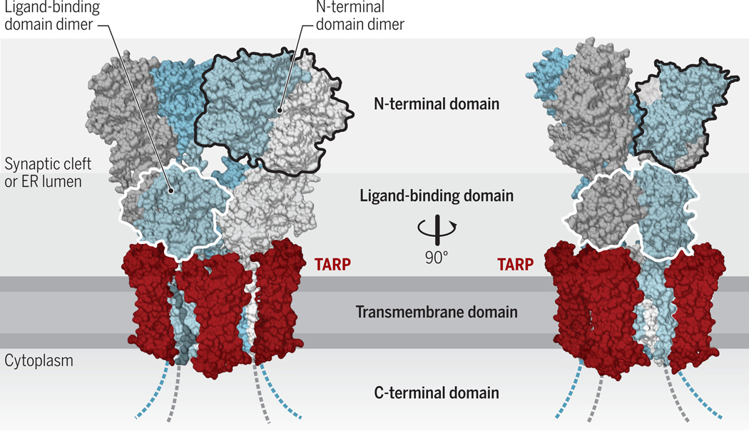
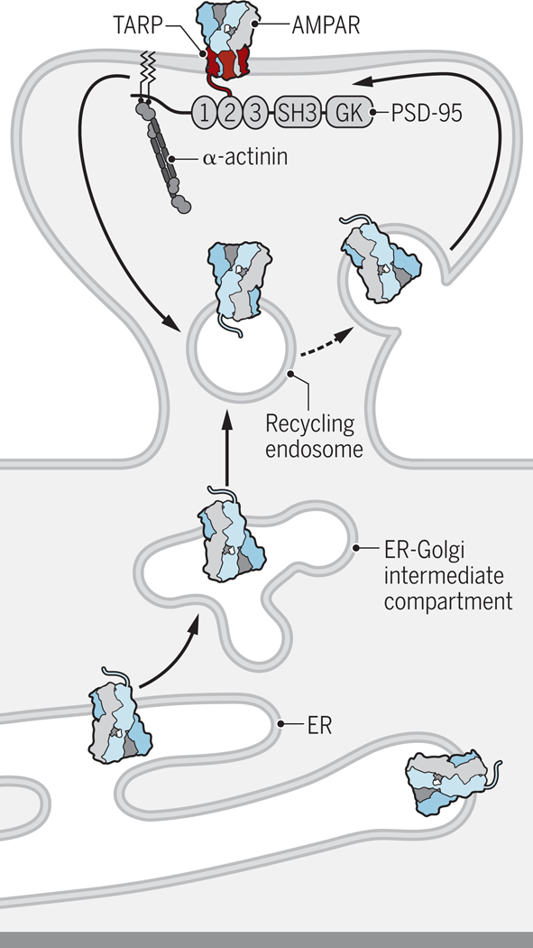
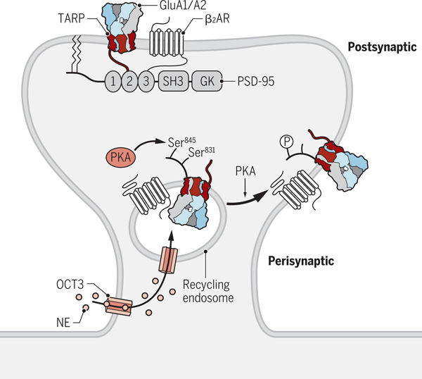
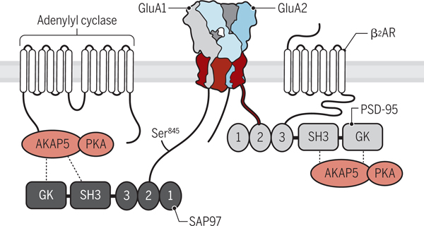
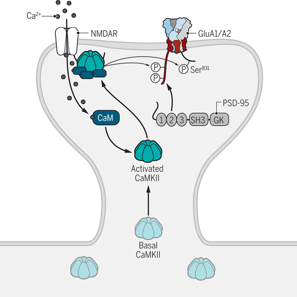
l-Glutamate is the main excitatory neurotransmitter in the brain, with postsynaptic responses to its release predominantly mediated by AMPA-type glutamate receptors (AMPARs). A critical component of synaptic plasticity involves changes in the number of responding postsynaptic receptors, which are dynamically recruited to and anchored at postsynaptic sites. Emerging findings continue to shed new light on molecular mechanisms that mediate AMPAR postsynaptic trafficking and localization. Accordingly, unconventional secretory trafficking of AMPARs occurs in dendrites, from the endoplasmic reticulum (ER) through the ER-Golgi intermediary compartment directly to recycling endosomes, independent of the Golgi apparatus. Upon exocytosis, AMPARs diffuse in the plasma membrane to reach the postsynaptic site, where they are trapped to contribute to transmission. This trapping occurs through a combination of both intracellular interactions, such as TARP (transmembrane AMPAR regulatory protein) binding to α-actinin-stabilized PSD-95, and extracellular interactions through the receptor amino-terminal domain. These anchoring mechanisms may facilitate precise receptor positioning with respect to glutamate release sites to enable efficient synaptic transmission.


Figures

Figure 1.
Figure 1.. Structural architecture of AMPARs.
AMPARs are formed by four subunits, which are conformationally (and functionally) distinct (‘pore-proximal’ subunits are in grey and ‘pore-distal subunits’ in blue). These subunits consist of an extracellular N-terminal domain, the ligand binding domain, an integral membrane domain, and an intracellular C-terminus domain, and form tetrameric receptors (chains A-D). The large extracellular region faces the ER-lumen during receptor biogenesis and ultimately projects into the synaptic cleft. The transmembrane AMPAR regulatory proteins (TARPs) interact with the receptor at up to four positions around the transmembrane domain (two non-equivalent positions indicated in red; structure reproduced from PDB:5WEO). Credit: Adapted by A. Kitterman/Science Signaling
Figure 2.
Figure 2.. Dendritic AMPAR trafficking.
AMPARs are synthesized either in the soma (not depicted) or dendritic shaft in the endoplasmic reticulum (ER). From the dendritic ER AMPARs traffic through the ER–Golgi intermediate compartment to recycling endosomes, which mediate surface insertion of AMPARs (31). It is unclear where exactly exocytosis occurs but it is likely either in the dendritic shaft near dendritic spines or in dendritic spines outside the postsynaptic density (PSD). AMPARs then move through lateral diffusion to the PSD, where they are trapped by PSD-95 and its homologues through their binding to the C-termini of TARPs. PSD-95 is anchored at postsynaptic sites by α-actinin. When and where TARPs, which are mostly if not exclusively translated in the soma (31), associate with AMPARs and especially those synthesized in dendrites is unknown. Credit: Kellie Holoski/Science Signaling
Figure 3.
Figure 3.. Regulation of perisynaptic AMPAR trafficking.
We propose that norepinephrine (NE) is shuttled by the amino acid transporter OCT3 localized in the plasma membrane from the cell exterior into the cytosol and then by OCT3 localized in recycling endosomes into their lumen. Here, NE stimulates the β2 adrenergic receptor (β2AR) associated with GluA1, which induces PKA activation and phosphorylation of Ser845 in GluA1. This phosphorylation event increases surface delivery of AMPARs from recycling endosomes. Lateral diffusion allows AMPARs to reach the PSD, where they are trapped by binding of the C_termini of TARPs to PSD-95. Credit: Kellie Holoski/Science Signaling
Figure 4.
Figure 4.. The AMPAR–β2 adrenergic receptor signaling complex.
The β2 adrenergic receptor binds through its extreme C-terminus to the third PDZ domain of PSD-95. In turn, the first two PDZ domains of PSD-95 bind to the C-termini of TARPs (red) including γ2 and γ8. Adenylyl cyclase binds through its N-terminus to the N-terminus of AKAP5 (also known as AKAP79 in humans, AKAP75 in cow, and AKAP150 in rodents), which binds through its C-terminus to PKA. AKAP5 is connected to AMPARs through SAP97, which binds to the C-terminus of GluA1, and potentially also through PSD-95. How Gs is linked to the β2 adrenergic receptor (β2AR) –AMPAR complex is unknown but could be through pre-association with the β2 adrenergic receptor. Credit: Kellie Holoski/Science Signaling
Figure 5.
Figure 5.. Regulation of postsynaptic AMPAR trafficking.
LTP-inducing stimuli trigger the influx of Ca2+ through NMDARs. Ca2+ binds to CaM and stimulates the activity of CaMKII. CaMKII is then recruited to the NMDAR complex by binding to the C-terminus of the GluN2B subunit. It subsequently phosphorylates the C-termini of TARPs including γ2 and γ8, which may lead to AMPAR trapping at the PSD. Phosphorylation of GluA1 on Ser831 by CaMKII also augments its channel activity. Credit: Kellie Holoski/Science Signaling

Similar articles

Cited by

References

    1. Micheva KD, Busse B, Weiler NC, O’Rourke N, Smith SJ, Single-synapse analysis of a diverse synapse population: proteomic imaging methods and markers. Neuron 68, 639–653 (2010). - PMC - PubMed
    1. Lu W et al., Subunit composition of synaptic AMPA receptors revealed by a single-cell genetic approach. Neuron 62, 254–268 (2009). - PMC - PubMed
    1. Renner MC et al., Synaptic plasticity through activation of GluA3-containing AMPA-receptors. eLife 6, (2017). - PMC - PubMed
    1. Traynelis SF et al., Glutamate receptor ion channels: structure, regulation, and function. Pharmacol Rev 62, 405–496 (2010). - PMC - PubMed
    1. Greger IH, Watson JF, Cull-Candy SG, Structural and Functional Architecture of AMPA-Type Glutamate Receptors and Their Auxiliary Proteins. Neuron 94, 713–730 (2017). - PubMed

Publication types