. 2019 Sep;22(3):381-393.
doi: 10.1007/s40477-018-0347-9. Epub 2019 Jan 1.

Pediatric cystic diseases of the kidney

Affiliations

Pediatric cystic diseases of the kidney

Federica Ferro et al. J Ultrasound. 2019 Sep.

Abstract

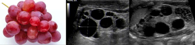
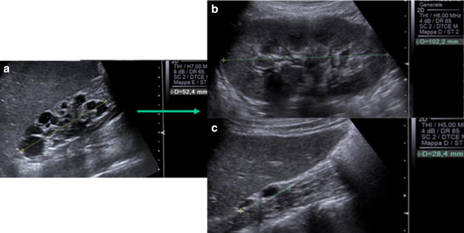
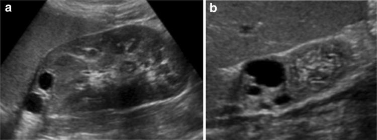
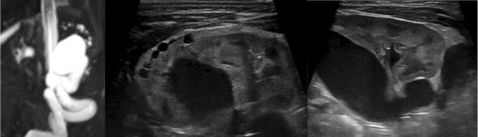
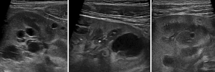
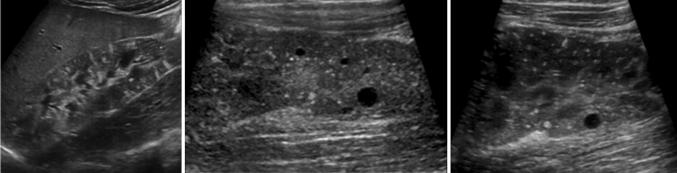
Pediatric renal cystic diseases include a variety of hereditary or non-hereditary conditions. Numerous classifications exist and new data are continuously published. Ultrasound is the primary technique for evaluating kidneys in children: conventional and high-resolution US allows a detailed visualization of renal parenchyma and of number, size and location of the cysts, hence representing the most important diagnostic imaging technique for the first diagnosis and follow-up of these young patients. The purpose of this pictorial essay is to review the spectrum of renal cystic lesions in children from simple, complex or malignant single cysts to the several poly/multicystic kidney diseases.

La patologia cistica renale in età pediatrica è costituita da svariate condizioni ereditarie e non. La letteratura propone diverse classificazioni, in continua evoluzione. L’ecografia rappresenta la prima metodica per la valutazione del rene nel neonato e nel bambino: l’esame con metodica tradizionale e con sonde ad alta risoluzione consente uno studio dettagliato del parenchima renale e delle cisti per numero, dimensioni e localizzazione. L’ecografia è pertanto il cardine nell’iter diagnostico e nel follow-up di questi piccoli pazienti. Obiettivo di questo lavoro è rivedere le peculiarità delle lesioni cistiche renali in età pediatrica: dalla cisti singola, semplice o complessa, allo spettro delle patologie multi/policistiche.

Keywords: Cystic renal disease; Kidney; Neonatal; Pediatric; Ultrasound.


Conflict of interest statement

Author Federica Ferro declares that she has no conflict of interest. Author Norberto Vezzali declares that he has no conflict of interest. Author Evi Comploj declares that she has no conflict of interest. Author Elena Pedron declares that she has no conflict of interest. Author Marco Di Serafino declares that he has no conflict of interest. Author Francesco Esposito declares that he has no conflict of interest. Author Piernicola Pelliccia declares that he has no conflict of interest. Author Eugenio Rossi declares that he has no conflict of interest. Author Massimo Zeccolini declares that he has no conflict of interest. Author Gianfranco Vallone declares that he has no conflict of interest.

Figures

Fig. 1
Fig. 1
Normal kidney (a) and two different aspects of dysplastic kidney (b, c)
Fig. 2
Fig. 2
Examples of various cortico-medullary differentiations in cystic renal diseases
Fig. 3
Fig. 3
Different localization of cysts: subcortical (a), cortico-medullary (b) and medullary (c)
Fig. 4
Fig. 4
Simple cysts: US and color Doppler typical aspects
Fig. 5
Fig. 5
Caliceal diverticulum: anechoic round lesion at the upper pole of the kidney with fortuitous evidence of a connection with a calyx (yellow double arrow)
Fig. 6
Fig. 6
Venous and delayed CT phases (a): presence of iodinated urine inside the ‘cystic lesion’ allows the diagnosis of caliceal diverticulum at the upper pole. Delayed MR phase of two different patients (b): a simple cyst homogenously hypodense and a caliceal diverticulum partially filled with enhancing urine (white arrow)
Fig. 7
Fig. 7
Bosniak III–IV cystic lesions: wall thickening, parietal nodule and a septum with nodule
Fig. 8
Fig. 8
A septate renal cyst at US evaluation and its simple appearance at multiphasic CT
Fig. 9
Fig. 9
Two examples of cystic pediatric renal tumors: US and MR appearance
Fig. 10
Fig. 10
MCDK: multiple cysts of variable size and dysplastic residual parenchyma in a central position
Fig. 11
Fig. 11
MCDK: US scan at diagnosis (a), check scans demonstrating compensatory hypertrophy of contralateral kidney (b) and involution of the affected kidney (c)
Fig. 12
Fig. 12
Two cases of segmental MCDK associated with duplicated collecting system: isolated multicystic dysplasia in upper pole (a) and multicystic dysplasia in upper pole associated with dysplastic kidney (b)
Fig. 13
Fig. 13
Acquired renal cysts. Patient with left-sided duplicated collecting system and ureteral ectopic insertion in vagina: coronal T2w image demonstrates severe upper ureteral dilatation associated with multiple tiny cysts with peripheral distribution at upper moiety. US depicts hydronephrosis and thinned dysplastic renal parenchyma with small peripheral cysts
Fig. 14
Fig. 14
Acquired renal cysts. Retrograde cystography demonstrates bilateral refluent megaureter. US depicts severe pyelic dilatation and thinned dysplastic hyperechoic renal parenchyma with small peripheral cysts
Fig. 15
Fig. 15
ARPKD: anteroposterior x-ray depicts pulmonary hypo/dysplasia, centralization of bowel associated with bulging flanks due to enlarged kidneys. US confirms nephromegaly, and high-resolution US allows the identification of linear cystic cortico-medullary microdilatations
Fig. 16
Fig. 16
High-frequency US in ARPKD: multiple hyperechogenic dots with “comet-tail” reverb and some macroscopic cysts between countless tiny microcysts in a radial distribution representing dilated tubules
Fig. 17
Fig. 17
US image of the liver in a child affected by ARPKD reveals hepatopathy and Caroli disease with extensive cystic biliary dilatation
Fig. 18
Fig. 18
ADPKD: small cortical and medullary cysts overlapping parenchyma with normal US appearance
Fig. 19
Fig. 19
NPHP: US depicts diffusely increased echogenicity of the renal parenchyma with loss/poor cortico-medullary differentiation, cysts, if present, affect the medulla as well as the cortico-medullary junction
Fig. 20
Fig. 20
Tuberous sclerosis: US shows combination of multiple hyperechoic angiomyolipomas and isolated small cysts

References

    1. Riccabona M, Ring E, Avni F. Imaging in renal agenesis, dysplasia, hypoplasia, and cystic diseases of the kidney. In: Riccabona M, editor. Pediatric urogenital radiology. Berlin: Springer; 2018. pp. 553–577.
    1. Gupta I, Bitzan M (2104) Cystic renal disease. In: Manual of pediatric nephrology. Springer, Berlin, pp 249–262
    1. Paliwalla M, Park K. A practical guide to urinary tract ultrasound in a child: pearls and pitfalls. Ultrasound. 2014;22:213–222. doi: 10.1177/1742271X14549795. - DOI - PMC - PubMed
    1. Chung E, Conran R, Schroder JW, Rohena-Quinquilla I, Rooks V. Pediatric polycystic kidney disease and other ciliopathies: radiologic-pathologic correlation. Radiographics. 2014;34:155–178. doi: 10.1148/rg.341135179. - DOI - PubMed
    1. MacRae K, Chisthi A, et al. Cystic renal disease. In: Chisti AS, et al., editors. Kidney and urinary tract disease in the newborn. Berlin: Springer; 2014. pp. 263–274.