. 2019;14(2):1561107.
doi: 10.1080/15592324.2018.1561107. Epub 2019 Jan 2.

Fluence rate dependence of red light-induced phosphorylation of plasma membrane H+-ATPase in stomatal guard cells

Affiliations

Fluence rate dependence of red light-induced phosphorylation of plasma membrane H+-ATPase in stomatal guard cells

Eigo Ando et al. Plant Signal Behav. 2019.

Abstract

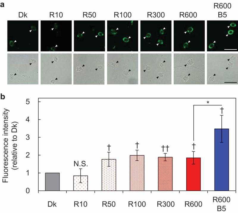
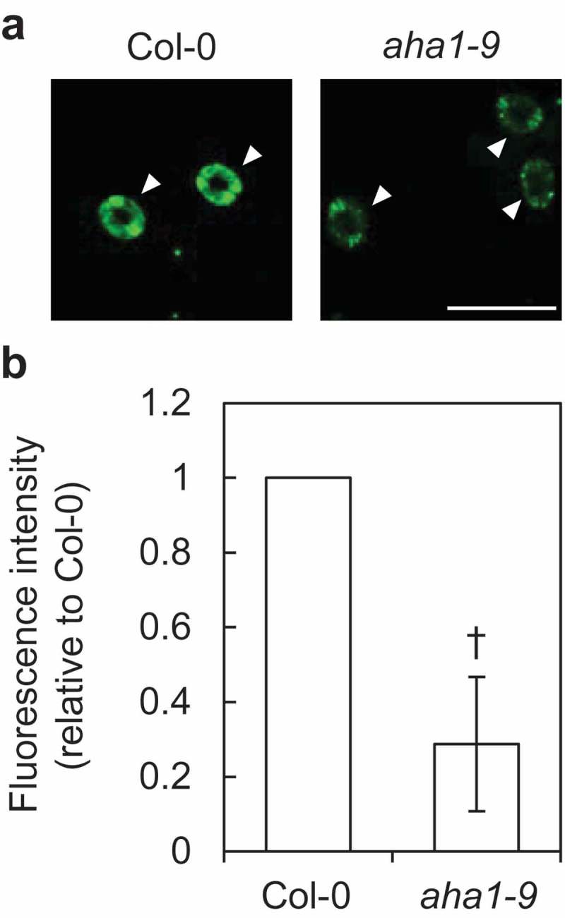
Stomatal opening is induced by red light as well as blue light. Recently, we established an immunohistochemical technique using whole leaves to study plasma membrane (PM) H+-ATPase in guard cells, which is an important enzyme driving stomatal opening. Our technique revealed that red light illuminated to whole leaves induces photosynthesis-dependent phosphorylation of C-terminal penultimate residue of PM H+-ATPase, threonine, in guard cells, which has been considered to be important for activation of PM H+-ATPase, and we proposed that red light promotes stomatal opening via activation of PM H+-ATPase in guard cells in whole leaves. Here, using our new immunohistochemical technique, we investigated fluence rate dependence of red light-induced phosphorylation of PM H+-ATPase. We found that illumination of red light at 50 µmol m-2 s-1, which was suggested to initiate photosynthesis, saturates phosphorylation of PM H+-ATPase. Furthermore, we immunohistochemically confirmed decrease in the amount of PM H+-ATPase protein in a knock-out mutant of AHA1, an isogene encoding the major isoform of PM H+-ATPase in guard cells, implying the importance of AHA1 as the major PM H+-ATPase protein in guard cells for light-induced stomatal opening.

Keywords: guard cells; immunohistochemistry; phosphorylation; photosynthesis; plasma membrane H-ATPase; red light.


Figures

Figure 1.
Figure 1.
Fluence rate dependence of red light-induced phosphorylation of PM H+-ATPase in guard cells. Mature leaves were harvested from dark-adapted plants and kept in the dark (Dk) or illuminated with the red light at 10, 50, 100, 300, and 600 µmol m–2 s–1 (R10, R50, R100, R300, and R600, respectively) for 30 min. As a positive control, leaves illuminated with red light (600 µmol m–2 s–1) for 30 min followed by blue light (5 µmol m–2 s–1) superimposed on red light for 2.5 min (R600 B5) were examined. (a) Typical fluorescent (upper panel) and bright field (lower panel) images are shown. Arrowheads indicate guard cells. Scale bars represent 50 µm. (b) Quantification of the fluorescence intensities was calculated as previously described19. Data represent means of relative fluorescence intensities obtained from three biologically independent measurements with standard deviations (SDs). Daggers indicate that the mean is statistically significantly higher than Dk set to 1.0. N.S., not significant (one-tailed one-sample t test; P < 0.05, ††P < 0.01, N.S., P = 0.72). Asterisk indicates that blue light-induced increase in the phosphorylation level of PM H+-ATPase is statistically significant (one-tailed Student’s t test; *P < 0.05).
Figure 2.
Figure 2.
Immunohistochemical detection of guard-cell PM H+-ATPase in wild-type Col-0 and aha1-9. (a) Typical fluorescent images are shown. Arrowheads indicate guard cells. Scale bar represents 50 µm. (b) Quantification of the fluorescence intensities was calculated as previously described.19 Data represent a mean of relative fluorescence intensity obtained from three biologically independent measurements with SD. Dagger indicates that the mean is statistically significantly lower than Col-0 set to 1.0. (one-tailed one-sample t test; P < 0.05).

Similar articles

Cited by

References

    1. Shimazaki K, Doi M, Assmann SM, Kinoshita T.. Light regulation of stomatal movement. Annu Rev Plant Biol. 2007;58:219–247. doi:10.1146/annurev.arplant.57.032905.105434. - DOI - PubMed
    1. Inoue S, Kinoshita T.. Blue light regulation of stomatal opening and the plasma membrane H+-ATPase. Plant Physiol. 2017;174(2):531–538. doi:10.1104/pp.17.00166. - DOI - PMC - PubMed
    1. Kinoshita T, Shimazaki K. Blue light activates the plasma membrane H+-ATPase by phosphorylation of the C-terminus in stomatal guard cells. Embo J. 1999;18(20):5548–5558. doi:10.1093/emboj/18.20.5548. - DOI - PMC - PubMed
    1. Kinoshita T, Shimazaki K. Biochemical evidence for the requirement of 14-3-3 protein binding in activation of the guard-cell plasma membrane H+-ATPase by blue light. Plant Cell Physiol. 2002;43(11):1359–1365. doi:10.1093/pcp/pcf167. - DOI - PubMed
    1. Toda Y, Wang Y, Takahashi A, Kawai Y, Tada Y, Yamaji N, Ma JF, Ashikari M, Kinoshita T. Oryza sativa H+-ATPase (OSA) is involved in the regulation of dumbbell-shaped guard cells of rice. Plant Cell Physiol. 2016;57(6):1220–1230. doi:10.1093/pcp/pcw070. - DOI - PMC - PubMed

Publication types

MeSH terms

Substances

LinkOut - more resources