. 2019 Feb;130(2):247-262.
doi: 10.1097/ALN.0000000000002529.

Nitric Oxide Donor Prevents Neonatal Isoflurane-induced Impairments in Synaptic Plasticity and Memory

Affiliations

Nitric Oxide Donor Prevents Neonatal Isoflurane-induced Impairments in Synaptic Plasticity and Memory

Michele L Schaefer et al. Anesthesiology. 2019 Feb.

Abstract

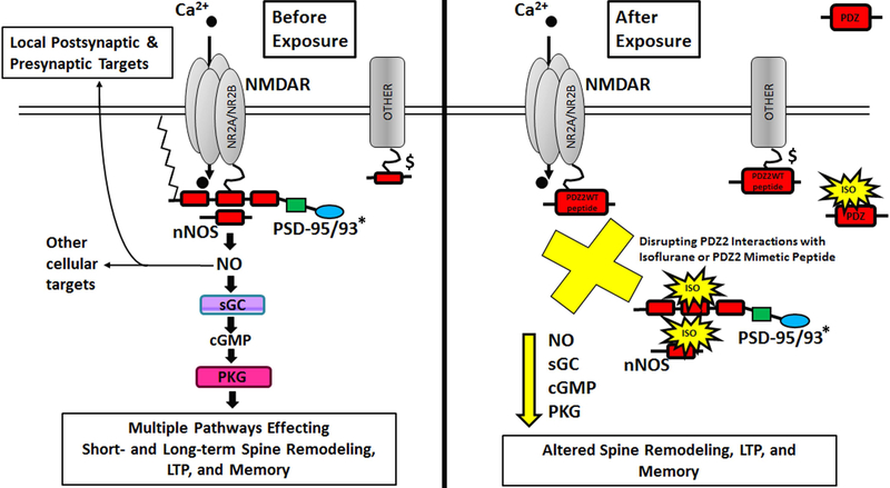
What we already know about this topic: Some general anesthetics have been shown to have adverse effects on neuronal development that affect neural function and cognitive behavior.Clinically relevant concentrations of inhalational anesthetics inhibit the postsynaptic density (PSD)-95, discs large homolog, and zona occludens-1 (PDZ) domain-mediated protein-protein interaction between PSD-95 or PSD-93 and N-methyl-D-aspartate receptors or neuronal NO synthase.

What this article tells us that is new: Neonatal PSD-95 PDZ2WT peptide treatment mimics the effects of isoflurane (~1 minimum alveolar concentration) by altering dendritic spine morphology, neural plasticity, and memory without inducing detectable increases in apoptosis or changes in synaptic density.These results indicate that a single dose of isoflurane (~1 minimum alveolar concentration) or PSD-95 PDZ2WT peptide alters dendritic spine architecture and functions important for cognition in the developing brain. This impairment can be prevented by administration of the NO donor molsidomine.

Background: In humans, multiple early exposures to procedures requiring anesthesia constitute a significant risk factor for development of learning disabilities and disorders of attention. In animal studies, newborns exposed to anesthetics develop long-term deficits in cognition. Previously, our laboratory showed that postsynaptic density (PSD)-95, discs large homolog, and zona occludens-1 (PDZ) domains may serve as a molecular target for inhaled anesthetics. This study investigated a role for PDZ interactions in spine development, plasticity, and memory as a potential mechanism for early anesthetic exposure-produced cognitive impairment.

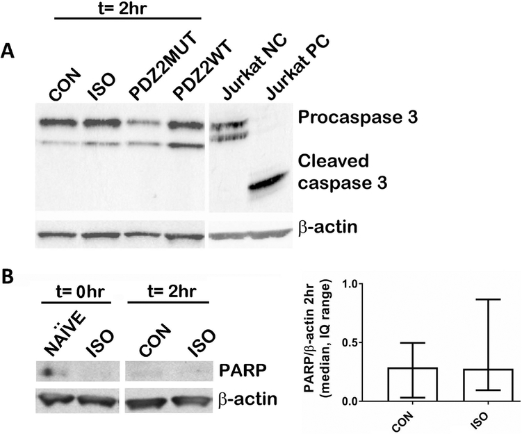
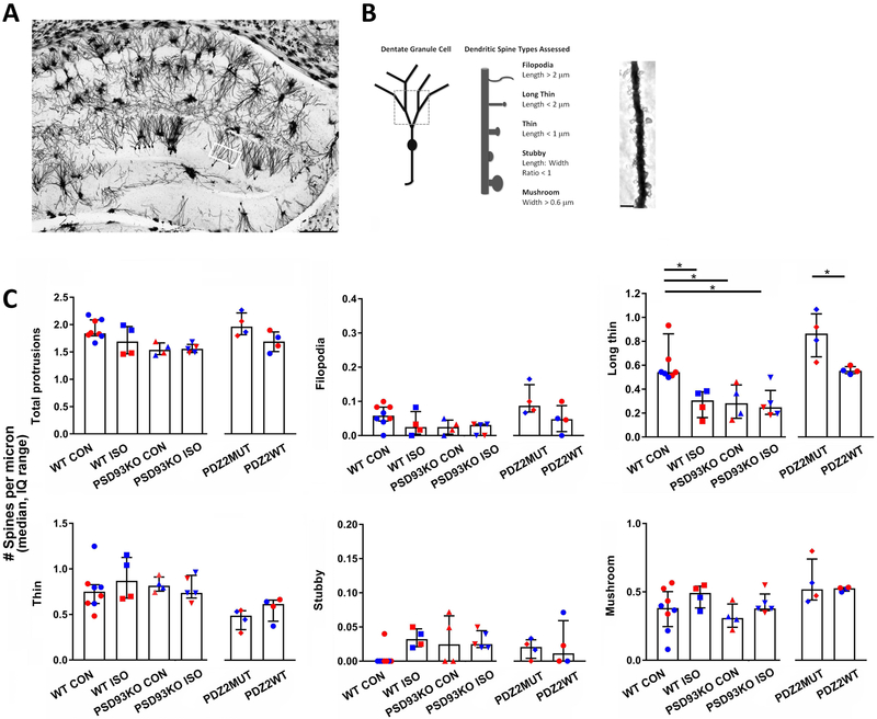
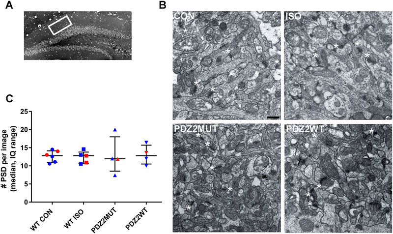
Methods: Postnatal day 7 mice were exposed to 1.5% isoflurane for 4 h or injected with 8 mg/kg active PSD-95 PDZ2WT peptide. Apoptosis, hippocampal dendritic spine changes, synapse density, long-term potentiation, and cognition functions were evaluated (n = 4 to 18).

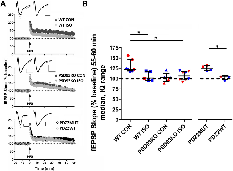
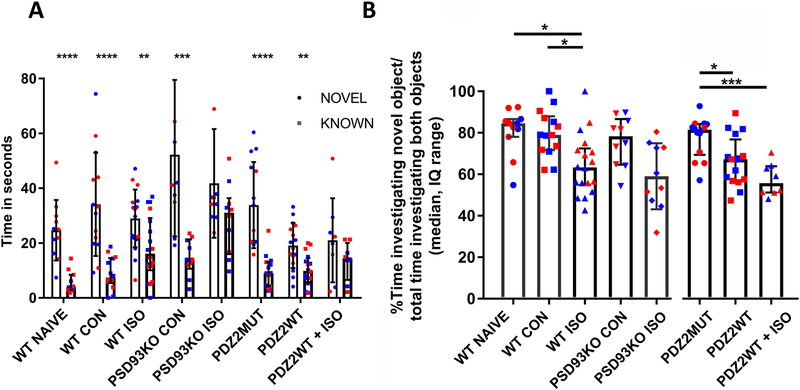
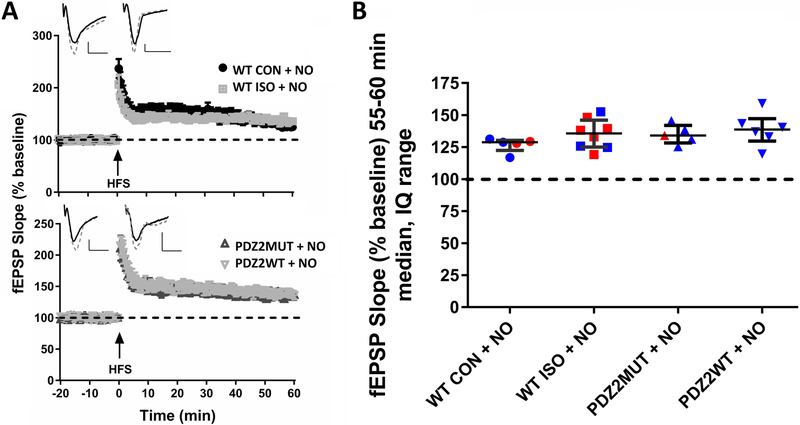
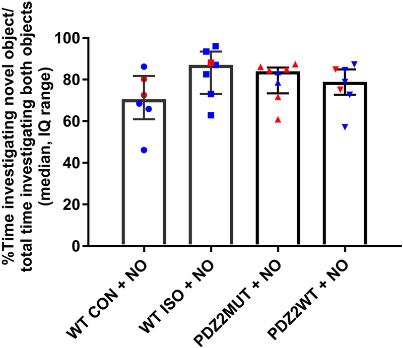
Results: Exposure of postnatal day 7 mice to isoflurane or PSD-95 PDZ2WT peptide causes a reduction in long thin spines (median, interquartile range [IQR]: wild type control [0.54, 0.52 to 0.86] vs. wild type isoflurane [0.31, 0.16 to 0.38], P = 0.034 and PDZ2MUT [0.86, 0.67 to 1.0] vs. PDZ2WT [0.55, 0.53 to 0.59], P = 0.028), impairment in long-term potentiation (median, IQR: wild type control [123, 119 to 147] and wild type isoflurane [101, 96 to 118], P = 0.049 and PDZ2MUT [125, 119 to 131] and PDZ2WT [104, 97 to 107], P = 0.029), and deficits in acute object recognition (median, IQR: wild type control [79, 72 to 88] vs. wild type isoflurane [63, 55 to 72], P = 0.044 and PDZ2MUT [81, 69 to 84] vs. PDZ2WT [67, 57 to 77], P = 0.039) at postnatal day 21 without inducing detectable differences in apoptosis or changes in synaptic density. Impairments in recognition memory and long-term potentiation were preventable by introduction of a NO donor.

Conclusions: Early disruption of PDZ domain-mediated protein-protein interactions alters spine morphology, synaptic function, and memory. These results support a role for PDZ interactions in early anesthetic exposure-produced cognitive impairment. Prevention of recognition memory and long-term potentiation deficits with a NO donor supports a role for the N-methyl-D-aspartate receptor/PSD-95/neuronal NO synthase pathway in mediating these aspects of isoflurane-induced cognitive impairment.


Conflict of interest statement

Conflicts of Interest: The authors declare no competing interests.

Figures

Fig. 1
Fig. 1
Diagram illustrating dissociation of NMDAR-PSD95/93*-nNOS interaction by ISO or PDZ2WT (active) peptide. *For simplicity, not all PSD-95 family members are represented. Before Exposure, The NMDA receptor is linked to downstream molecules such as nNOS through PSD-95: through its first and second PDZ domain, PSD-95 forms a ternary complex by binding to both the tSXV motif of NMDAR NR2 subunit and to the PDZ domain in nNOS . Disrupting NMDAR-PSD-95/93-nNOS complexes can reduce the efficiency by which calcium ions activate the signaling molecule nNOS. After Exposure, This disruption is achieved by exposure to inhalational anesthetics, or the intracellular introduction of PDZ2WT peptide ; this is expected to bind to NMDAR NR2 . $ Inhalational anesthetics (and presumably PDZ2WT peptide) can also inhibit interactions between PSD-95 PDZ2 domain and Shaker-type potassium channel Kv1.4 as well as other excitatory receptor channels related to anesthesia and other proteins not shown here for simplicity .
Fig. 2
Fig. 2
Western blot assays did not show any significant differences in apoptosis in our exposure paradigm. PND7 mice were exposed for 4-hours and harvested 2-hours after cessation of exposure. A, Western blot analysis for total caspase-3 revealed the presence of procaspase 3 but did not reveal any detectable cleaved caspase-3 in mice exposed to CON (O2), ISO (1.5% ISO in O2), PDZ2MUT (8 mg/kg inactive peptide), PDZ2WT (8 mg/kg active peptide). Negative and positive Jurkat control proteins were run to demonstrate sensitivity of the caspase-3 antibody. B, Western blot analysis for PARP did not reveal significant differences between CON (O2) and ISO (1.5% ISO in O2). Left, representative blot. Right, Densitometry data are plotted as median, interquartile range. CON (n=6) vs ISO (n=6), p=0.818. Data were analyzed using Mann Whitney test. Gender was not determined.
Fig. 3
Fig. 3
Neonatal exposure to ISO or PDZ2WT peptide alters hippocampal dendritic spine morphology and development. A, dorsal hippocampal region of a Golgi preparation illustrating dendrites from the superior blade of the dentate gyrus (DG) subregion of interest (white box); scale bar represents 250 μm. B, schematics showing dendrite branches and spine types sampled and a representative dendritic segment with spines; scale bar represents 2.5 μm. Spines were assessed on dendritic segments distal to the first and second branch points. C, Distribution of dendritic spines according to morphological type in DG among exposed groups assessed at PND21. WT and PSD-93 KO PND7 mice were exposed to O2 (control, 100% O2) or ISO (1.5% ISO in O2); WT PND7 mice were also injected with PDZ2MUT (8 mg/kg inactive peptide) or PDZ2WT (8 mg/kg active peptide). (WT CON = 8, WT ISO = 4, PSD93KO CON = 4, PSD93KO ISO = 5, PDZ2MUT = 4, PDZ2WT = 4). Data from individual animals are plotted and color coded by gender (red=female and blue=male). Data are plotted as median number of spines per micron with interquartile range). Data were analyzed with Kruskal-Wallis followed by Dunn’s multiple comparison correction and Mann-Whitney tests. A *p < 0.05 was considered significant.
Fig. 4
Fig. 4
Neonatal exposure to ISO or PDZ2WT peptide did not have an acute impact on the number of hippocampal postsynaptic densities. A, dorsal hippocampal region of a semi-thin section illustrating DG subregion of interest (white box). B, representative ultrastructure images from PND21 mice exposed at PND7 to O2 (WT CON, 100% O2), WT ISO (1.5% ISO in O2), PDZ2MUT peptide (8 mg/kg inactive peptide), or PDZ2WT peptide (8 mg/kg active peptide); scale bar represents 500 nm. Asterisks showing some PSDs (not all are labeled). C, plots showing the median with interquartile range number of PSD’s. WT CON (n=6) vs WT ISO (n=5), p=0.829; PDZ2MUT (n=4) vs PDZ2WT (n=4), p=0.742. Data from individual animals are plotted and color coded by gender (red=female and blue=male). Data were analyzed with Mann-Whitney.
Fig. 5
Fig. 5
Neonatal exposure to ISO or PDZ2WT peptide impairs long-term potentiation (LTP) in hippocampal CA1 at PND21. A, high frequency stimulation (HFS) induced robust LTP in WT CON (top; WT CON, 100% O2) and inactive PDZ2MUT (bottom) treated groups in hippocampal Schafer collateral to CA1 pathway. ISO (top) and PDZ2WT (bottom) exposure as well as PSD93 deficiency (middle) impaired the expression of LTP. Example traces are shown in upper left quadrants of fEPSP plots. WT CON and WT ISO (top), PSD93KO CON and PSD93KO ISO (middle), and PDZ2MUT and PDZ2WT (bottom) treated groups at baseline before HFS (solid line trace) and the average of 55-60 min after HFS (dashed line trace). B, the median of normalized fEPSP 55-60min after HFS showed significant differences between WT CON (n=7) vs WT ISO (n=5), p=0.049 and PDZ2MUT (n=4) vs PDZ2WT (n=4), p=0.028 treated groups. Significant differences were not observed between WT CON vs PSD93KO CON (n=5), p=0.056 but were observed between WT CON vs PSD93KO ISO (n=8), p=0.025 groups. Data from individual animals are plotted and color coded by gender (red=female and blue=male). Data were analyzed with Kruskal-Wallis followed by post-hoc Dunn’s test and Mann Whitney tests. Values were considered significant at *p < 0.05 or less. Data were plotted as median and interquartile range. Scale bar: 10ms, 0.25mV.
Fig. 6.
Fig. 6.
Neonatal exposure to ISO or PDZ2WT peptide causes a subtle but significant decrease in acute recognition memory. A, plots showing percent of time animals spent investigating novel or known objects among experimental groups(WT Naïve, n=11, p<0.0001; WTCON, n=14, p<0.0001; WT ISO, n=18, p=0.005; PSD93KO CON, n=10, p=0.001; PDZ2MUT, n=14, p<0.0001; PDZ2WT, n=16, p=0.001). The double hit animals were unable to significantly discriminate between novel and known objects (PSD93KO ISO, n=10, p=0.098; PDZ2WT+ISO, n=8, p=0.227). Data were plotted as mean and SD. Data were analyzed with two-tailed t-tests known vs. novel. B, plots showing discrimination index as percent of time animals spent investigating novel object over the total time investigating novel and known objects multiplied by 100. ISO exposed WT animals have a subtle but significant decrement in recognition memory as compared to controls (WT NAÏVE vs WT ISO, p = 0.022; WT CON vs WT ISO, p = 0.043; WT NAÏVE vs WTCON, p >0.999). PSD93 deficiency did not have a significant effect on recognition memory (WT NAÏVE vs PSD93KO CON, p>0.999; WT CON vs PSD93KOCON, p>0.999). ISO exposed PSD93 KO animals were not significantly different from PSD93 KO controls (PSD93KO CON vs PSD93KO ISO, p=0.176). ISO exposed PSD93KO animals differed from WT controls (WT NAÏVE vs PSD93KO ISO, p=0.011; WT CON vs PSD93KO ISO, p=0.022). Active peptide exposed animals have a subtle but significant decrement in recognition memory as compared to inactive peptide controls (PDZ2MUT vs PDZ2WT, p=0.038; PDZ2MUT vs PDZ2WT + ISO, p<0.001) ISO exposure did not further significantly impair peptide exposed animals (PDZ2WT vs PDZ2WT + ISO, p=0.385). Data from individual animals are plotted and color coded by gender (red=female and blue=male). Data were analyzed with Kruskal-Wallis followed by post-hoc Dunn’s test. Data were plotted as median and interquartile range. *p < 0.05, **p < 0.01, ***p < 0.001, and ****p < 0.0001.
Fig.7.
Fig.7.
Treatment with NO donor prevents the negative effect of ISO and PDZ2WT peptide on hippocampal LTP. A, robust LTP was induced by HFS in all groups. In upper left quadrants example traces of WT CON+NO and WT ISO+NO (top), PDZ2MUT+NO and PDZ2WT+NO (bottom) treated groups before HFS (solid line trace) and 55-60min after HFS (dashed line trace) are shown. B, the median of normalized fEPSP 55-60min after HFS no longer shows a significant difference between WT CON and WT ISO when NO donor is added (WT CON + NO (n=5) vs ISO + NO (n=8), p=0.284) or between PDZ2MUT and PDZ2WT (PDZ2MUT + NO (n=5) vs PDZ2WT + NO (n=6), p=0.662). Data were analyzed with Mann-Whitney tests. Data from individual animals are plotted and color coded by gender (red=female and blue=male). Data were plotted as median and interquartile range. Values were considered significant at *p < 0.05 or less. Scale bar: 10ms, 0.25mV.
Fig. 8.
Fig. 8.
Treatment with NO donor prevents ISO or PDZ2WT peptide induced impairment in acute recognition memory. Plots showing discrimination index as percent of time animals spent investigating novel object over the total time investigating novel and known objects multiplied by 100. There is no longer a significant difference between WT CON and WT ISO when NO donor is added (WT CON + NO (n=6) vs ISO + NO (n=7), p=0.073) or between PDZ2MUT and PDZ2WT (PDZ2MUT + NO (n=8) vs PDZ2WT + NO (n=7), p=0.778). Data from individual animals are plotted and color coded by gender (red=female and blue=male). Data were plotted as median and interquartile range. Data were analyzed with Mann-Whitney tests. Values were considered significant at *p < 0.05 or less.

Similar articles

Cited by

References

    1. Jackson WM, Gray CD, Jiang D, Schaefer ML, Connor C, Mintz CD: Molecular Mechanisms of Anesthetic Neurotoxicity: A Review of the Current Literature. J Neurosurg Anesthesiol 2016, 28:361–72. - PMC - PubMed
    1. Gentry KR, Steele LM, Sedensky MM, Morgan PG: Early developmental exposure to volatile anesthetics causes behavioral defects in Caenorhabditis elegans. Anesth Analg 2013, 116:185–9. - PMC - PubMed
    1. Jevtovic-Todorovic V, Hartman RE, Izumi Y, Benshoff ND, Dikranian K, Zorumski CF, Olney JW, Wozniak DF: Early exposure to common anesthetic agents causes widespread neurodegeneration in the developing rat brain and persistent learning deficits. J Neurosci 2003, 23:876–82. - PMC - PubMed
    1. Brambrink AM, Evers AS, Avidan MS, Farber NB, Smith DJ, Zhang X, Dissen GA, Creeley CE, Olney JW: Isoflurane-induced neuroapoptosis in the neonatal rhesus macaque brain. Anesthesiology 2010, 112:834–41. - PMC - PubMed
    1. Flick RP, Katusic SK, Colligan RC, Wilder RT, Voigt RG, Olson MD, Sprung J, Weaver AL, Schroeder DR, Warner DO: Cognitive and behavioral outcomes after early exposure to anesthesia and surgery. Pediatrics 2011, 128:e1053–61. - PMC - PubMed

Publication types