

A Rapid Protocol for Intraoperative Assessment of Peripheral Nerve Myelinated Axon Count and Its Application to Cross-Facial Nerve Grafting
- PMID: 30601328
- PMCID: PMC7147971
- DOI: 10.1097/PRS.0000000000005338
A Rapid Protocol for Intraoperative Assessment of Peripheral Nerve Myelinated Axon Count and Its Application to Cross-Facial Nerve Grafting
Erratum in
-
A Rapid Protocol for Intraoperative Assessment of Peripheral Nerve Myelinated Axon Count and Its Application to Cross-Facial Nerve Grafting: Correction.Plast Reconstr Surg. 2020 May;145(5):1341. doi: 10.1097/PRS.0000000000007007. Plast Reconstr Surg. 2020. PMID: 32332563 No abstract available.
Abstract
Background: Donor nerve myelinated axon counts correlate with functional outcomes in reanimation procedures; however, there exists no reliable means for their intraoperative quantification. In this article, the authors report a novel protocol for rapid quantification of myelinated axons from frozen sections, and demonstrate its applicability to surgical practice.
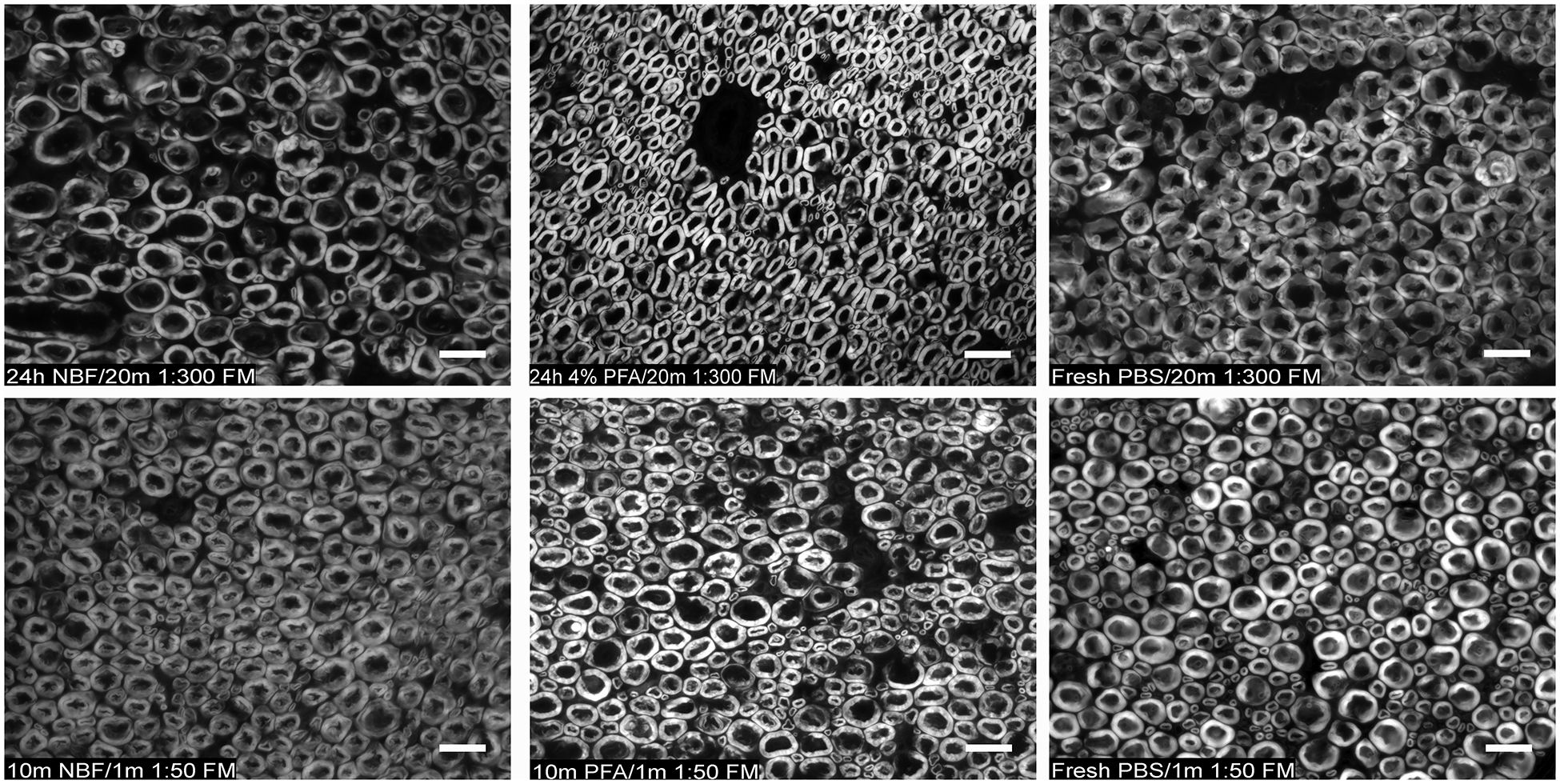
Methods: The impact of various fixation and FluoroMyelin Red staining strategies on resolved myelin sheath morphology from cryosections of rat and rabbit femoral and sciatic nerves was assessed. A protocol comprising fresh cryosection and rapid staining was developed, and histomorphometric results were compared against conventional osmium-postfixed, resin-embedded, toluidine blue-stained sections of rat sciatic nerve. The rapid protocol was applied for intraoperative quantification of donor nerve myelinated axon count in a cross-facial nerve grafting procedure.
Results: Resolution of myelinated axon morphology suitable for counting was realized within 10 minutes of tissue harvest. Although mean myelinated axon diameter appeared larger using the rapid fresh-frozen as compared to conventional nerve processing techniques (mean ± SD; rapid, 9.25 ± 0.62 μm; conventional, 6.05 ± 0.71 μm; p < 0.001), no difference in axon counts was observed on high-power fields (rapid, 429.42 ± 49.32; conventional, 460.32 ± 69.96; p = 0.277). Whole nerve myelinated axon counts using the rapid protocol herein (8435.12 ± 1329.72) were similar to prior reports using conventional osmium processing of rat sciatic nerve.
Conclusions: A rapid protocol for quantification of myelinated axon counts from peripheral nerves using widely available equipment and techniques has been described, rendering possible intraoperative assessment of donor nerve suitability for reanimation.
Conflict of interest statement
Figures


References
-
- Harii K, Ohmori K, Torii S. Free gracilis muscle transplantation, with microneurovascular anastomoses for the treatment of facial paralysis. A preliminary report. Plast Reconstr Surg. 1976;57(2):133–143. - PubMed
-
- O’Brien BM, Franklin JD, Morrison WA. Cross-facial nerve grafts and microneurovascular free muscle transfer for long established facial palsy. Br J Plast Surg. 1980;33(2):202–215. - PubMed
-
- Klebuc M Masseter–to-Facial Nerve Transfer: A new technique for facial reanimation. J Reconstr Microsurg. 2006;22(03):A101.
-
- Eisenhardt SU, Eisenhardt NA, Thiele JR, Stark GB, Bannasch H. Salvage procedures after failed facial reanimation surgery using the masseteric nerve as the motor nerve for free functional gracilis muscle transfer. JAMA facial plastic surgery. 2014;16(5):359–363. - PubMed
-
- Bae YC, Zuker RM, Manktelow RT, Wade S. A comparison of commissure excursion following gracilis muscle transplantation for facial paralysis using a cross-face nerve graft versus the motor nerve to the masseter nerve. Plast Reconstr Surg. 2006;117(7):2407–2413. - PubMed
Publication types
MeSH terms
Substances
Grants and funding
LinkOut - more resources
Full Text Sources
Other Literature Sources
