Review
. 2019 Jun 20:88:691-724.
doi: 10.1146/annurev-biochem-013118-111153. Epub 2019 Jan 11.

Mechanism and Regulation of Centriole and Cilium Biogenesis

Affiliations
Review

Mechanism and Regulation of Centriole and Cilium Biogenesis

David K Breslow et al. Annu Rev Biochem. .

Abstract

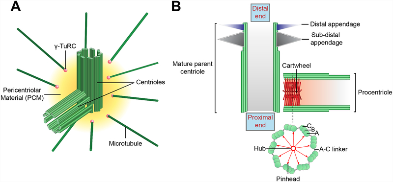
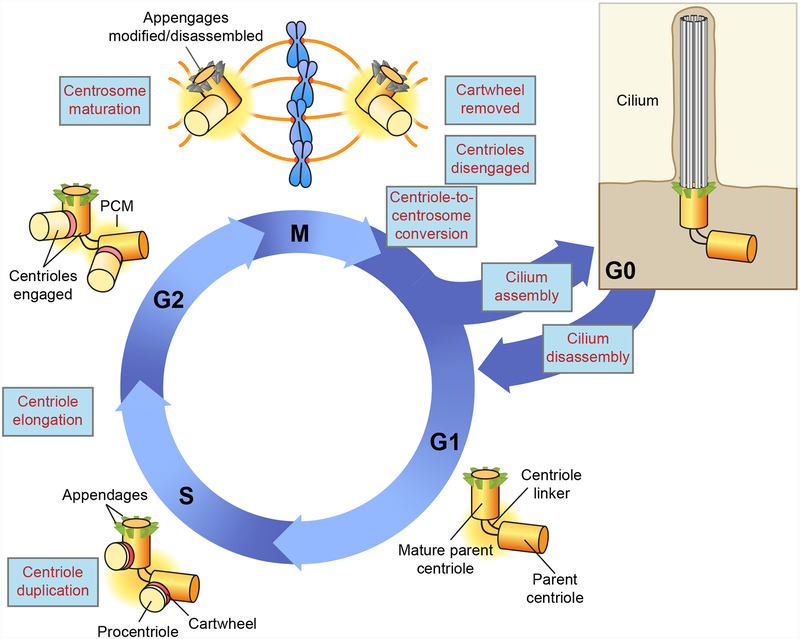
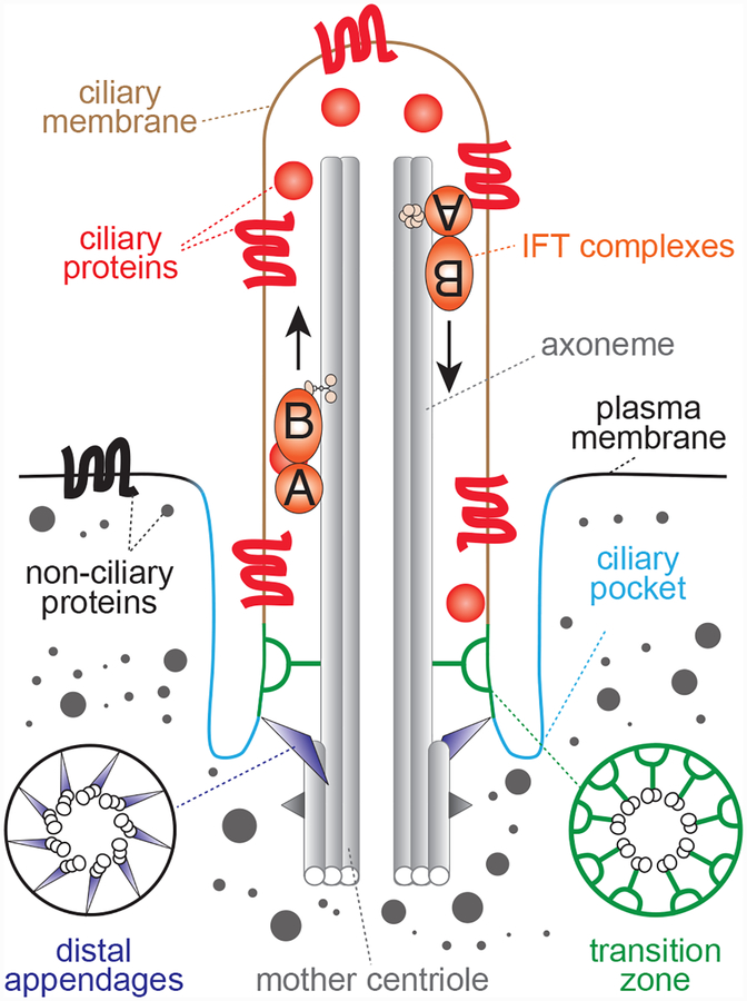
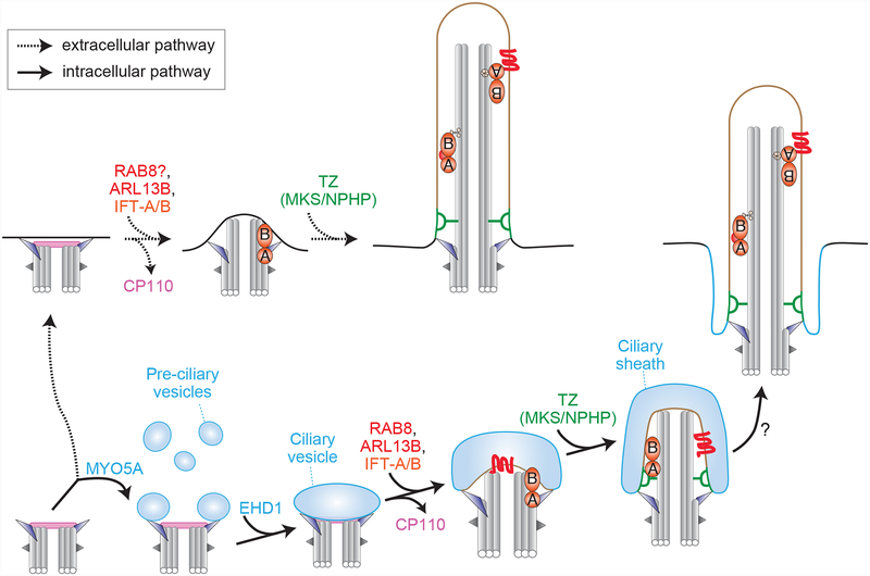
The centriole is an ancient microtubule-based organelle with a conserved nine-fold symmetry. Centrioles form the core of centrosomes, which organize the interphase microtubule cytoskeleton of most animal cells and form the poles of the mitotic spindle. Centrioles can also be modified to form basal bodies, which template the formation of cilia and play central roles in cellular signaling, fluid movement, and locomotion. In this review, we discuss developments in our understanding of the biogenesis of centrioles and cilia and the regulatory controls that govern their structure and number. We also discuss how defects in these processes contribute to a spectrum of human diseases and how new technologies have expanded our understanding of centriole and cilium biology, revealing exciting avenues for future exploration.

Keywords: cell cycle; centriole; centrosome; cilia; ciliopathy; mitosis.


Figures

Figure 1.
Figure 1.. Centriole and centrosome structure
A) Architecture of the mammalian centrosome. The centrosome is comprised of a pair of orthogonally oriented centrioles surrounded by a proteinaceous Pericentriolar Material (PCM). The PCM contains proteins required for microtubule nucleation and anchoring, such as the γ-Tubulin Ring Complex (γTuRC) (pink spheres). B) Schematic illustration of a mature parent centriole and associated procentriole. Centrioles are cylindrical strictures comprised of nine triplet microtubules, each of which contains a complete A-tubule and an incomplete B and C-tubule. The cartwheel is present in the proximal lumen of the procentriole and is formed by a central hub from which nine spokes emanate. Each spoke terminates in a pinhead structure that binds to the A-tubule of the microtubule triplet. The A-tubule of one triplet is linked to the C-tubule of the adjacent triplet via an A-C linker. Mature parent centrioles are decorated at their distal end with ninefold symmetric distal and sub-distal appendages.
Figure 2.
Figure 2.. Regulation of centriole and cilium biogenesis during the cell cycle
G1 cells contain two parent centrioles connected at their base by a flexible linker. At the beginning of S phase, each parent centriole assembles one new procentriole aligned orthogonal to its proximal end. This arrangement is termed ‘engagement’ and acts to prevent the reduplication of the parent centriole. The procentrioles elongate as cells progress through the cycle and in late G2, the flexible linker that holds the two parent centrioles together is dissolved to permit centrosome separation. In preparation for mitotic spindle formation, centrosome maturation occurs resulting in PCM expansion. During mitosis the cartwheel is removed from the lumen of the procentriole. At the end of mitosis, the centriole pair disengages and loses its orthogonal arrangement. This step is required to relicense the parent centriole for duplication in the next cell cycle. At the same time the procentriole is converted into a parent centriole. This ‘centriole-to-centrosome’ conversion allows the procentriole to recruit PCM material and acquire competence for duplication. The distal and sub-distal appendages are transiently modified/disassembled in mitosis. In G1 appendages form on the mature parent centriole that was created one and half cell cycle earlier. In quiescent cells, the mature parent centriole can migrate to the plasma membrane and initiate the formation of the axoneme of a cilium. Cell cycle re-entry is accompanied by the disassembly of the cilium.
Figure 3.
Figure 3.. Primary cilium structure
Architecture of a mammalian primary cilium, highlighting key structural features. The axonemal microtubules form the core of the cilium and extend from the mature parent centriole, which is docked at the plasma membrane. This docking is mediated by the mature centriole’s distal appendages and often occurs at a site on the cell surface where the plasma membrane is invaginated. This invaginated region of the plasma membrane adjacent to the cilium is known as the ciliary pocket is a key site for exo/endocytosis of ciliary materials. Although the cilium lacks a delimiting membrane, it contains a distinct complement of soluble and membrane proteins. This compartmentalization is enabled by diffusion barriers near the base of the cilium at a region known as the transition zone (TZ). The transition zone is made up of several functional and physical modules, including MKS and NPHP proteins, which are mutated in Meckel Syndrome and Nephronophthisis, respectively. Selective trafficking of proteins to cilia across the transition zone is mediated by trafficking machineries, such as IFT-A and IFT-B, that cooperate with ciliary kinesin and dynein motors. Additionally, IFT-A and IFT-B mediate protein transport within cilia along the axonemal microtubules and are required for ciliogenesis.
Figure 4.
Figure 4.. Pathways for primary cilium assembly
The mature parent centriole (bottom left) serves as the foundation for primary cilium assembly via either an ‘extracellular’ pathway (top, dashed arrows) or an intracellular pathway (bottom, solid arrows). In the intracellular pathway, key steps include i) MYO5A-dependent recruitment of pre-ciliary vesicles to the distal appendages, ii) EHD1-mediated fusion of these vesicles to form an enlarged ciliary vesicle, iii) the growth of the ciliary vesicle via the joint action of RAB8, ARL13B, and the IFT complexes, a process that occurs in conjunction with removal of the CP110 cap from the distal end of the fully mature centriole, iv) the growth of the axoneme, formation of the transition zone, and maturation of the ciliary vesicle into distinct domains corresponding to the ciliary sheath and the nascent ciliary membrane, and v) the fusion of the ciliary sheath with the ciliary membrane, which exposes the cilium to the external environment. In the extracellular pathway, a key distinction is that the mature parent centriole initially migrates to the cell surface and docks to the plasma membrane via its distal appendages. Subsequent steps appear to occur in a similar fashion as the intracellular pathway, although the precise sequence of events and molecular requirements are not fully knowns.
Figure 5.
Figure 5.. Application of functional screening to study cilia and centrioles
A) Overview of pooled functional screening using CRISPR/Cas9. A pool of sgRNAs is introduced into Cas9-expressing cells by lentiviral transduction. Transduced cells can then be grown under conditions that select for a functional property of centrioles or cilia or in the absence of such selection (note that the cells in question may need to engineered such that centrioles/cilia control a selectable phenotype). Deep sequencing is then used to analyze the composition of sgRNAs present at the outset of the experiment (T0 – e.g. the sgRNA library used to make lentiviral particles), in the unselected pool at the end of the experiment (Tend unselected), and in the selected pool at the end of the experiments (Tend selected). If sgRNAs targeting a particular gene are consistently depleted (or enriched) in the final selected sample relative to the final unselected sample, then the gene in question regulates centriole or cilium function. Similarly, changes in sgRNA abundance between the T0 sample and the final unselected sample reveal genes that affect cell growth. B) Schematic illustration of using growth phenotype screens conducted in different cell lines (indicated by cells of different shape) to identify genes with shared function. Hierarchical clustering of growth phenotypes across all cell lines can be used to group and identify genes having a shared function. C) Several centriolar genes, including members of the TED complex (bold labels), exhibit highly correlated patterns of growth phenotypes to that of C14orf80/TEDC1 across 436 cell lines in the Achilles dataset (Avana public 18Q2). The growth phenotypes for knockout of C14orf80/TEDC1 were compared to those for all other genes in the dataset, yielding the plotted distribution of correlation coefficients. Correlation values between TEDC1 and other TED complex components are indicated.

Similar articles

Cited by

References

    1. Carvalho-Santos Z, Azimzadeh J, Pereira-Leal JB, Bettencourt-Dias M. 2011. Evolution: Tracing the origins of centrioles, cilia, and flagella. J Cell Biol 194: 165–75 - PMC - PubMed
    1. Pickett-Heaps JD. 1971. The autonomy of the centriole: fact or fallacy? Cytobios 3: 205–14
    1. Gonczy P 2012. Towards a molecular architecture of centriole assembly. Nat Rev Mol Cell Biol 13: 425–35 - PubMed
    1. Greenan GA, Keszthelyi B, Vale RD, Agard DA. 2018. Insights into centriole biogenesis and evolution revealed by cryoTomography of doublet and triplet centrioles. bioRxiv - PMC - PubMed
    1. Wang JT, Stearns T. 2018. The ABCs of Centriole Architecture: The Form and Function of Triplet Microtubules. Cold Spring Harb Symp Quant Biol - PMC - PubMed

Publication types

LinkOut - more resources