

Mitochondrial dysfunction in type 2 diabetes mellitus: an organ-based analysis
- PMID: 30601700
- PMCID: PMC6397358
- DOI: 10.1152/ajpendo.00314.2018
Mitochondrial dysfunction in type 2 diabetes mellitus: an organ-based analysis
Abstract
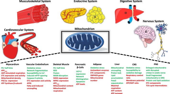
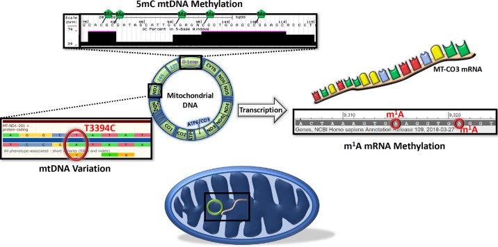
Type 2 diabetes mellitus (T2DM) is a systemic disease characterized by hyperglycemia, hyperlipidemia, and organismic insulin resistance. This pathological shift in both circulating fuel levels and energy substrate utilization by central and peripheral tissues contributes to mitochondrial dysfunction across organ systems. The mitochondrion lies at the intersection of critical cellular pathways such as energy substrate metabolism, reactive oxygen species (ROS) generation, and apoptosis. It is the disequilibrium of these processes in T2DM that results in downstream deficits in vital functions, including hepatocyte metabolism, cardiac output, skeletal muscle contraction, β-cell insulin production, and neuronal health. Although mitochondria are known to be susceptible to a variety of genetic and environmental insults, the accumulation of mitochondrial DNA (mtDNA) mutations and mtDNA copy number depletion is helping to explain the prevalence of mitochondrial-related diseases such as T2DM. Recent work has uncovered novel mitochondrial biology implicated in disease progressions such as mtDNA heteroplasmy, noncoding RNA (ncRNA), epigenetic modification of the mitochondrial genome, and epitranscriptomic regulation of the mtDNA-encoded mitochondrial transcriptome. The goal of this review is to highlight mitochondrial dysfunction observed throughout major organ systems in the context of T2DM and to present new ideas for future research directions based on novel experimental and technological innovations in mitochondrial biology. Finally, the field of mitochondria-targeted therapeutics is discussed, with an emphasis on novel therapeutic strategies to restore mitochondrial homeostasis in the setting of T2DM.
Keywords: diabetes mellitus; mitochondria dysfunction.
Conflict of interest statement
No conflicts of interest, financial or otherwise, are declared by the authors.
Figures
References
-
- Akude E, Zherebitskaya E, Chowdhury SK, Smith DR, Dobrowsky RT, Fernyhough P. Diminished superoxide generation is associated with respiratory chain dysfunction and changes in the mitochondrial proteome of sensory neurons from diabetic rats. Diabetes 60: 288–297, 2011. doi:10.2337/db10-0818. - DOI - PMC - PubMed
-
- Anderson EJ, Lustig ME, Boyle KE, Woodlief TL, Kane DA, Lin CT, Price JW III, Kang L, Rabinovitch PS, Szeto HH, Houmard JA, Cortright RN, Wasserman DH, Neufer PD. Mitochondrial H2O2 emission and cellular redox state link excess fat intake to insulin resistance in both rodents and humans. J Clin Invest 119: 573–581, 2009. doi:10.1172/JCI37048. - DOI - PMC - PubMed
Publication types
MeSH terms
Substances
Grants and funding
LinkOut - more resources
Full Text Sources
Medical