. 2019 Mar 1;30(5):579-590.
doi: 10.1091/mbc.E18-11-0718. Epub 2019 Jan 2.

Clathrin plaques and associated actin anchor intermediate filaments in skeletal muscle

Affiliations

Clathrin plaques and associated actin anchor intermediate filaments in skeletal muscle

Agathe Franck et al. Mol Biol Cell. .

Abstract

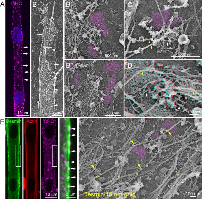
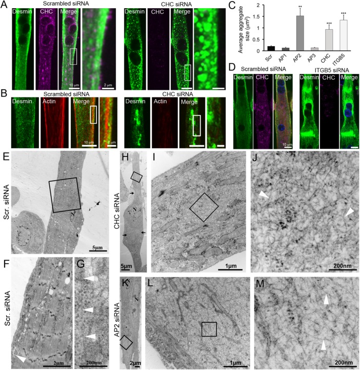
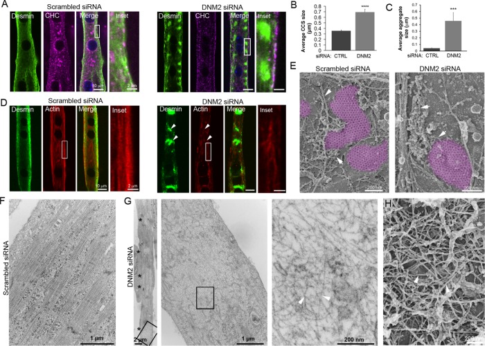
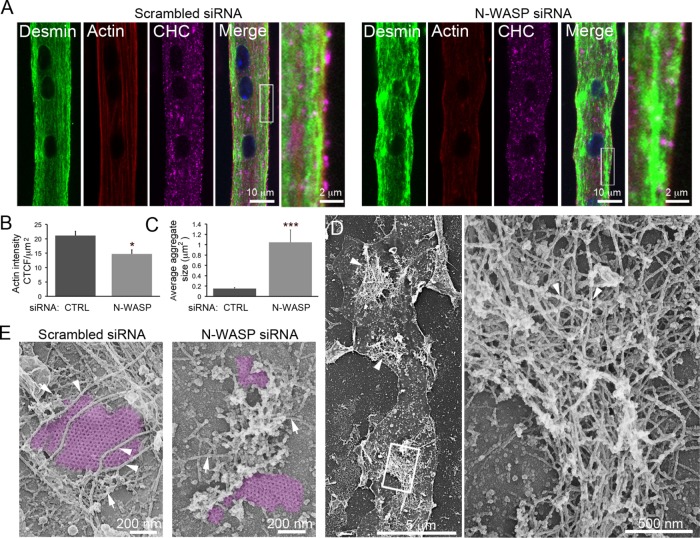
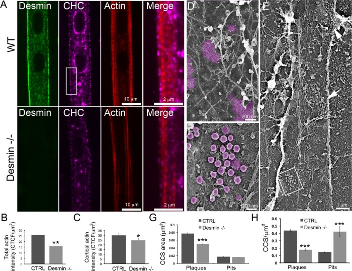
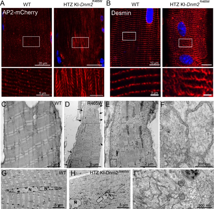
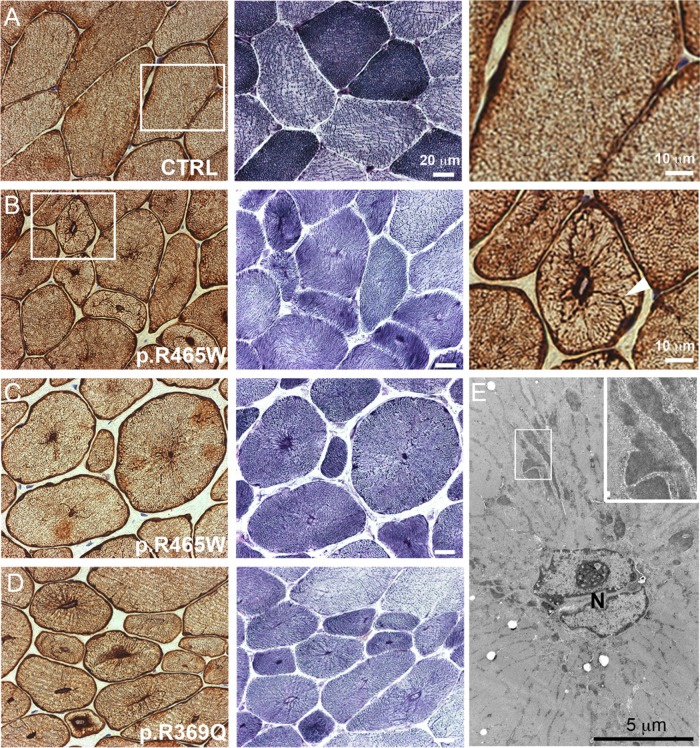
Clathrin plaques are stable features of the plasma membrane observed in several cell types. They are abundant in muscle, where they localize at costameres that link the contractile apparatus to the sarcolemma and connect the sarcolemma to the basal lamina. Here, we show that clathrin plaques and surrounding branched actin filaments form microdomains that anchor a three-dimensional desmin intermediate filament (IF) web. Depletion of clathrin plaque and branched actin components causes accumulation of desmin tangles in the cytoplasm. We show that dynamin 2, whose mutations cause centronuclear myopathy (CNM), regulates both clathrin plaques and surrounding branched actin filaments, while CNM-causing mutations lead to desmin disorganization in a CNM mouse model and patient biopsies. Our results suggest a novel paradigm in cell biology, wherein clathrin plaques act as platforms capable of recruiting branched cortical actin, which in turn anchors IFs, both essential for striated muscle formation and function.


Figures

FIGURE 1:
FIGURE 1:
Clathrin-coated plaques anchor desmin IFs. (A) Immunofluorescent staining of CHC (magenta) in differentiated mouse primary myotubes. (B) Survey view of an unroofed primary mouse myotube. (B′, B″) Higher-magnification views corresponding to the boxed regions in B. (C, D) Higher-magnification views of clathrin plaques and associated cytoskeletal structures in unroofed control primary myotubes. Intermediate filaments are indicated with yellow arrowheads and actin filaments are indicated using arrows. For D, use glasses for 3D viewing of anaglyph (left eye = red). (E) Immunofluorescent staining of desmin (green), actin (red), and CHC (magenta) in mouse primary myotubes. (F) Higher-magnification view of clathrin plaques and associated cytoskeletal structures in unroofed control primary myotubes immunolabeled using a primary antibody against desmin and secondary antibodies coupled to 15 nm gold beads. Images are representative of at least four independent experiments. Gold beads are pseudocolored in yellow, and some are indicated with yellow arrowheads. Clathrin-coated structures are highlighted in purple.
FIGURE 2:
FIGURE 2:
Clathrin-coated plaques are required for desmin IF organization. (A) Immunofluorescent staining of desmin (green) and CHC (magenta) in mouse primary myotubes treated with control or CHC siRNA. Images are representative of at least 10 independent experiments. (B) Desmin (green) and actin staining (red) in mouse primary myotubes treated with control or CHC siRNA. (C) Average desmin aggregate size in myotubes treated with control siRNA or siRNA against CHC, AP1, AP2, AP3, or b5 integrin (ITGB5) (n = 30–50 myotubes). Data presented as mean ± SEM; **, p < 0.01, ***, p < 0.001 using a two-tailed Student’s t test). (D) Immunofluorescent staining of desmin (green) and CHC (magenta) in mouse primary myotubes treated with control or ITGB5 siRNA. (E–L) Thin-section EM of primary myotubes treated with control (E–G), CHC (H–J), or AP2 (K–M) siRNA. I and L are higher-magnification views of IF tangles from H and K, respectively. Images are representative of at least two to four independent experiments. IFs are indicated with arrowheads.
FIGURE 3:
FIGURE 3:
DNM2 is required for desmin and actin organization around clathrin plaques. (A) Immunofluorescent staining of desmin (green) and actin (red) in mouse primary myotubes treated with control or DNM2 siRNA. Images are representative of at least seven independent experiments. (B) Average clathrin-coated structure (CCS) size in myotubes treated with control or siRNA against DNM2 (n = 20 myotubes). (C) Quantification of desmin aggregate fluorescence in myotubes treated with control or DNM2 siRNA (n = 20 myotubes). (D) Desmin (green) and actin staining (red) in mouse primary myotubes treated with control or DNM2 siRNA. (E) High-magnification view of unroofed primary mouse myotubes treated with control or DNM2 siRNA. Actin structures are indicated using arrows. Images are representative of at least 12 independent experiments. (F, G) Thin-section EM of extensively differentiated control (F) or DNM2-depleted (G) myotubes. Images are representative of at least eight independent experiments. Arrowheads denote bone fide IFs. (H) High-magnification view of a desmin aggregate from unroofed primary mouse myotubes treated with DNM2 siRNA. Data are presented as mean ± SEM; ***, p < 0.001, ****, p < 0.0001, using a two-tailed Student’s t test.
FIGURE 4:
FIGURE 4:
N-WASP is indispensable for desmin and actin organization around clathrin plaques. (A) Immunofluorescent staining of desmin (green), CHC (magenta), and actin (red) in mouse primary myotubes treated with control or N-WASP siRNA. Images are representative of at least five independent experiments. (B) Quantification of cortical actin fluorescence intensity in myotubes treated with control or N-WASP siRNA (n = 20 myotubes). (C) Quantification of desmin aggregate size in myotubes treated with control or N-WASP siRNA (n = 20 myotubes). (D) Survey view of desmin aggregates (arrowheads) from unroofed primary mouse myotubes treated with N-WASP siRNA. (E) High-magnification views of unroofed primary mouse myotubes treated with control or N-WASP siRNA. IFs are indicated with arrowheads, and actin structures are indicated using arrows. Images are representative of at least five independent experiments. Data are presented as mean ± SEM; *, p < 0.05, ***, p < 0.001, using a two-tailed Student’s t test.
FIGURE 5:
FIGURE 5:
Clathrin plaques and actin are altered in desmin knockout mice. (A) Immunofluorescence detection of desmin (green), CHC (magenta), and actin staining (phalloidin, red) in WT or desmin knockout (desmin−/−) mouse myotubes. Scale bars: 10 µm; 2 µm (insets). (B) Quantification of total actin fluorescence intensity in WT or desmin knockout−/− myotubes (n = 18–21 myotubes). (C) Quantification of cortical actin fluorescence intensity in WT or desmin knockout mice myotubes (n = 18–21 myotubes). (D) High-magnification view of an unroofed primary mouse myotube from WT mice. (E) Survey view of an unroofed desmin−/− myotube. Arrows indicate clathrin-coated pit accumulations. (F) Higher-magnification view corresponding to the boxed region in E. (G) Morphometric analysis of clathrin-coated structure size from WT or desmin knockout mouse myotubes. (H) Morphometric analysis of clathrin-coated structure density per membrane surface from WT or desmin−/− myotubes. Images are representative of at least five independent experiments. Data are presented as mean ± SEM; *, p < 0.05, **, p < 0.01, ***, p < 0.001, using a two-tailed Student’s t test.
FIGURE 6:
FIGURE 6:
Desmin IF defects in HTZ KI-Dnm2 R465W mice. (A) AP2 mCherry distribution between WT and HTZ KI-Dnm2R465W mice at the surface of myofibers on longitudinal muscle cryosections. (B) Confocal sections from the surface of WT (left panel) or HTZ KI-Dnm2 R465W (right panel) mouse dissociated skeletal muscle fibers immunolabeled with desmin antibody. Images are representative of at least three independent experiments. (C–F) Thin-section EM of surface longitudinal skeletal muscle sections from either WT (C) or HTZ KI-Dnm2 R465W (D–F) mice. The presence of desmin filament aggregates at the periphery of the fiber are clearly visible on the insets. (G–I) Longitudinal skeletal muscle sections from the core of either WT (G) or HTZ KI-Dnm2 R465W mice (H, I) displaying central nuclei. The presence of desmin filament aggregates around myonuclei is visible in the inset (I).
FIGURE 7:
FIGURE 7:
Desmin IF defects in human CNM patients. (A–D) Muscle biopsy sections of a control patient (A), two patients with p.R465W DNM2 mutation (B, C), and one patient with p.R369Q DNM2 mutation (D) were either immunohistochemically labeled against desmin or stained with NADH-TR reaction C. Muscle sections from the patient reveal a strong desmin labeling of radial sarcoplasmic strands. NADH-TR reaction C on muscle section from CNM patients displays a high percentage of small rounded fibers with centralized nuclei and few fibers with typical radiating sarcoplasmic strands. Radial desmin labeling is indicated with arrowheads in B. Note that desmin labeling continuously extends from the central nucleus to the sarcolemma. (E) Thin-section EM of muscle from a CNM patient with the p.R465W mutation presenting characteristic radial strands. Note the dilated Z-band material in the inset.

Similar articles

Cited by

References

    1. Bitoun M, Maugenre S, Jeannet PY, Lacène E, Ferrer X, Laforêt P, Martin JJ, Laporte J, Lochmüller H, Beggs AH, et al. (2005). Mutations in dynamin 2 cause dominant centronuclear myopathy. Nat Genet , 1207–1209. - PubMed
    1. Bonazzi M, Kühbacher A, Toledo-Arana A, Mallet A, Vasudevan L, Pizarro-Cerdá J, Brodsky FM, Cossart P. (2012). A common clathrin-mediated machinery co-ordinates cell-cell adhesion and bacterial internalization. Traffic , 1653–1666. - PMC - PubMed
    1. Brodsky FM. (2012). Diversity of clathrin function: new tricks for an old protein. Annu Rev Cell Dev Biol , 309–336. - PubMed
    1. Capetanaki Y, Papathanasiou S, Diokmetzidou A, Vatsellas G, Tsikitis M. (2015). Desmin related disease: a matter of cell survival failure. Curr Opin Cell Biol , 113–120. - PMC - PubMed
    1. Chorev DS, Moscovitz O, Geiger B, Sharon M. (2014). Regulation of focal adhesion formation by a vinculin-Arp2/3 hybrid complex. Nat Commun , 3758. - PubMed

Publication types

MeSH terms

LinkOut - more resources