. 2019 Jan 2;15(1):e1007882.
doi: 10.1371/journal.pgen.1007882. eCollection 2019 Jan.

Conserved function of the matriptase-prostasin proteolytic cascade during epithelial morphogenesis

Affiliations

Conserved function of the matriptase-prostasin proteolytic cascade during epithelial morphogenesis

Leonard Drees et al. PLoS Genet. .

Abstract

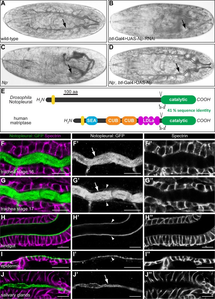
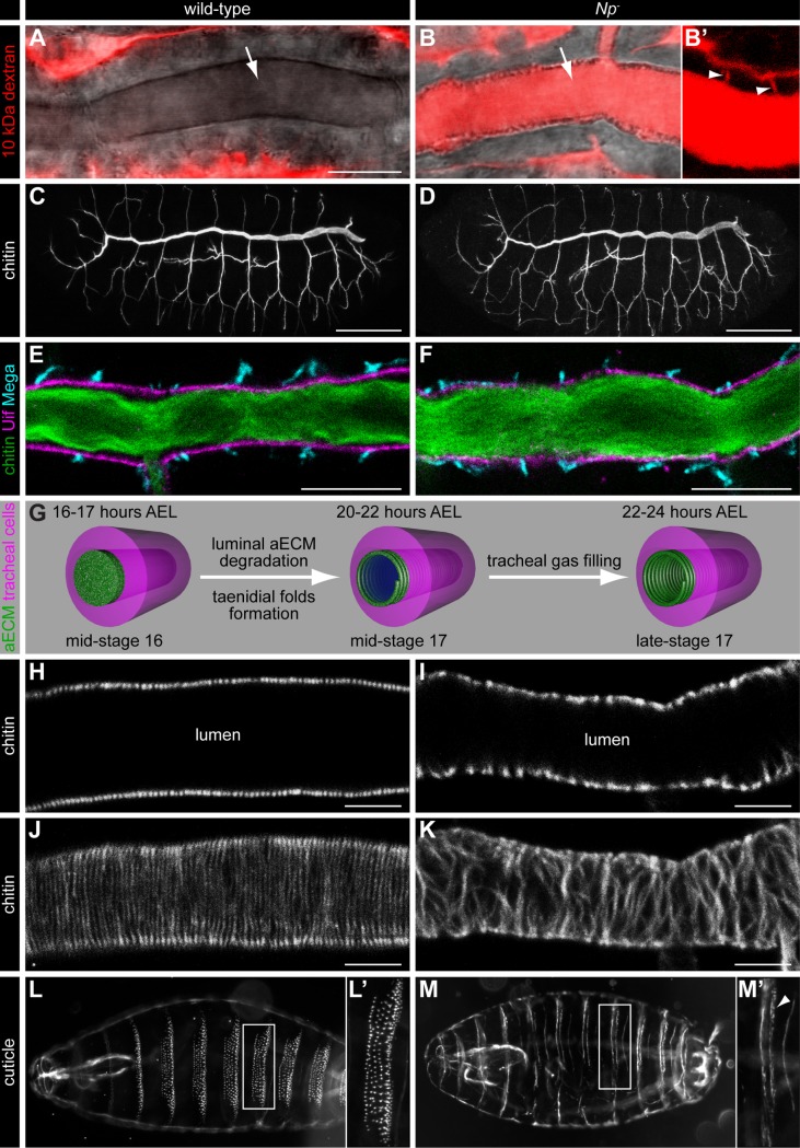
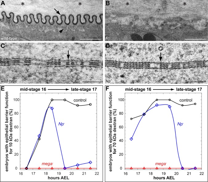
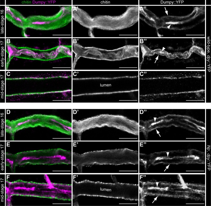
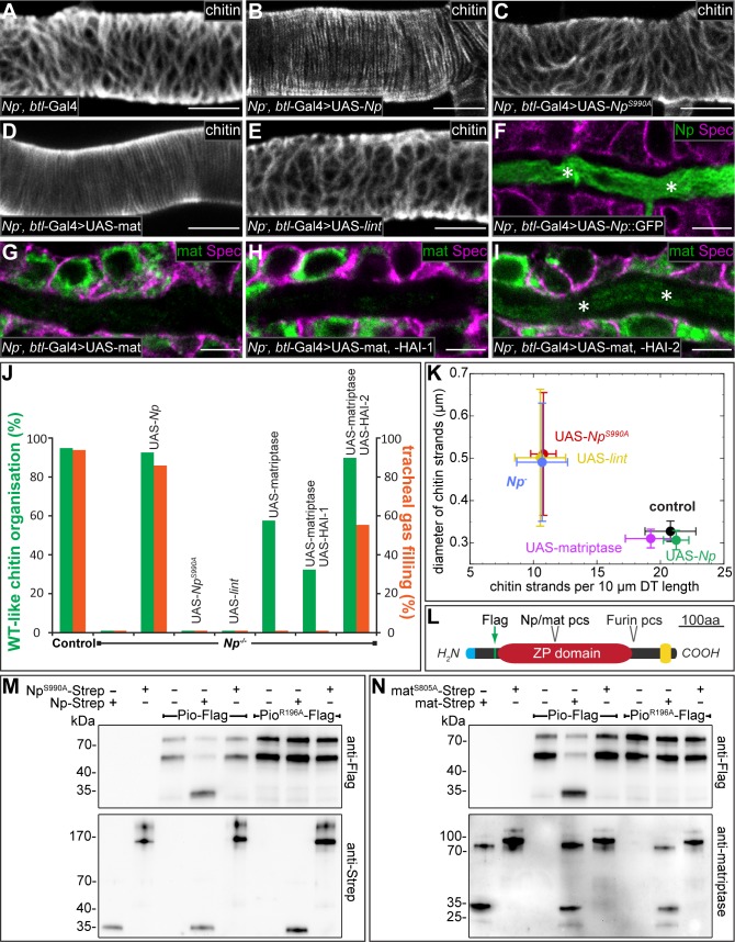
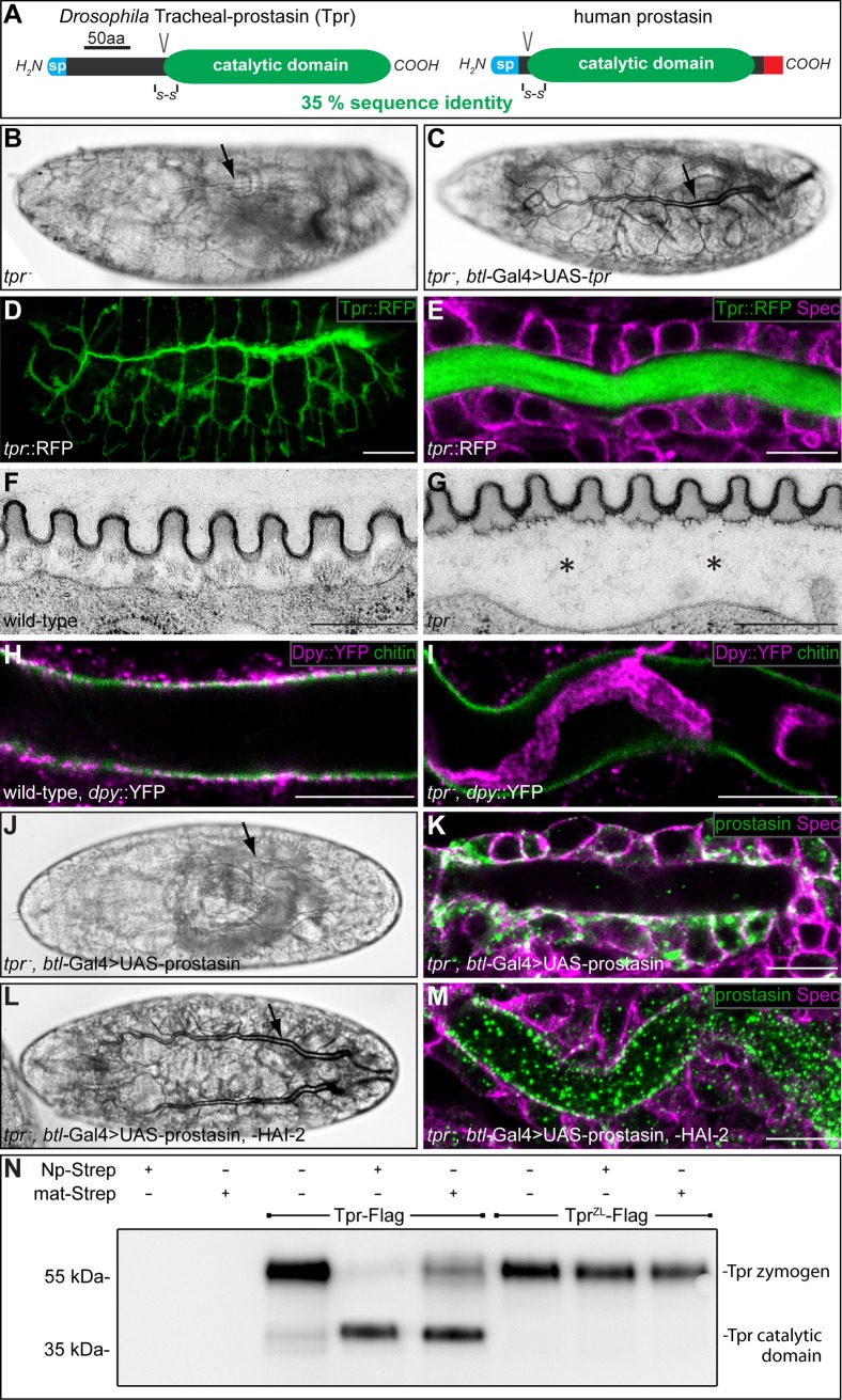
Extracellular matrix (ECM) assembly and remodelling is critical during development and organ morphogenesis. Dysregulation of ECM is implicated in many pathogenic conditions, including cancer. The type II transmembrane serine protease matriptase and the serine protease prostasin are key factors in a proteolytic cascade that regulates epithelial ECM differentiation during development in vertebrates. Here, we show by rescue experiments that the Drosophila proteases Notopleural (Np) and Tracheal-prostasin (Tpr) are functional homologues of matriptase and prostasin, respectively. Np mediates morphogenesis and remodelling of apical ECM during tracheal system development and is essential for maintenance of the transepithelial barrier function. Both Np and Tpr degrade the zona pellucida-domain (ZP-domain) protein Dumpy, a component of the transient tracheal apical ECM. Furthermore, we demonstrate that Tpr zymogen and the ZP domain of the ECM protein Piopio are cleaved by Np and matriptase in vitro. Our data indicate that the evolutionarily conserved ZP domain, present in many ECM proteins of vertebrates and invertebrates, is a novel target of the conserved matriptase-prostasin proteolytic cascade.


Conflict of interest statement

The authors have declared that no competing interests exist.

Figures

Fig 1
Fig 1. Notopleural is required for embryonic tracheal gas filling and encodes a serine protease related to human matriptase.
(A-D) Bright field light microscopic images of stage 17 wild-type (A), btl-Gal4; UAS-RNAi-GD13443 (B), NpP6/NpC2 mutant (C) and NpP6,btl-Gal4/NpC2,UAS-Np mutant (D) embryos. Wild-type embryos show gas filled tracheal tubes at the end of embryogenesis (arrow in A). RNAi-mediated tracheal knock-down of CG34350 (Np) leads to lack of tracheal gas filling (arrow in B). NpP6/NpC2 mutant embryos lack gas filling (arrow in C) while Np mutant embryos with tracheal expression of Np show normal gas filling of the tracheal system (arrow in D). (E) Schema showing the protein domain organisations of Drosophila Np and human matriptase. The transmembrane domains (yellow), the SEA (sea urchin sperm protein/enteropeptidase/agrin), CUB (Cls/Clr, urchin embryonic growth factor, bone morphogenetic protein-1), LDLa (low-density lipoprotein receptor class A) and the catalytic protease domains are shown. Conserved disulphide bridges (-S-S-) and zymogen activation cleavage sites (V) are indicated. (F-J”) Confocal LSM images of whole-mount antibody stainings of Np::GFP embryos at stage 16 (F-F”, H-J”) and stage 17 (G-G”) stained with anti-Spectrin (magenta) and anti-GFP (green, Np::GFP) antibodies. Np::GFP is expressed in the tracheal system (F-G”), the hindgut (H-H”), the epidermis (I-I”) and the salivary glands (J-J”). Np::GFP is localized in the tracheal lumen during stage 16 (arrow in F’) and 17 (arrow in G’) and localizes to the apical membrane of tracheal cells during stage 17 (arrowheads in G’). In the hindgut and epidermis, Np::GFP is localized exclusively at the apical cell membranes (arrowheads in H’ and I’). In the salivary glands, Np::GFP is localized exclusively in the lumen (arrow in J’). Scale bars correspond to 10 μm.
Fig 2
Fig 2. Notopleural is required for epithelial barrier function and aECM formation.
(A-B’) Confocal LSM images of tracheal dorsal trunks of wild-type (A) and NpP6/NpC2 mutant (B) stage 17 embryos after Texas Red-labelled 10 kDa dextran injection into the haemocoel. Texas Red dextran (red) is not found in the dorsal trunk lumen of wild-type embryos (arrow in A), but is detectable in the dorsal trunk lumen (arrow in B) and the tracheal paracellular space (arrowheads in B’) of NpP6/NpC2 mutant embryos. (B’) shows the tracheal dorsal trunk with increased contrast to visualize the paracellular space (arrowheads in B’). (C, D) Confocal LSM images of whole-mount stainings of stage 16 wild-type (C) and NpP6/NpC2 mutant (D) embryos with FITC labelled chitin-binding probe (CBP). CBP binds the luminal chitin matrix and outlines the tracheal network during embryogenesis. The tracheal network formation of Np mutant embryos (D) is indistinguishable from wild-type embryos (C). (E, F) Confocal LSM images of tracheal dorsal trunks of wild-type (E) and NpP6/NpC2 mutant (F) stage 16 embryos stained with CBP (green), anti-Uif (magenta) and anti-Mega (cyan) antibodies. (G) Schema of tracheal aECM maturation and liquid clearance. The aECM (green) in the tracheal lumen is degraded after mid-stage 16 and the mature taenidial folds (green spiral) form at the apical side of tracheal cells (magenta) during mid-stage 17. Liquid (blue) is cleared from the tracheal lumen during late-stage 17. Time data refer to embryonic development at 22°C. (H-K) Confocal LSM optical sections (H, I) and sagittal z-stack projection images (J, K) of dorsal trunks of wild-type (H, J) and NpP6/NpC2 mutant (I, K) stage 17 embryos stained with CBP. (L-M’) Dark field microscopy images of stage 17 wild-type (L, L’) and NpP6/NpC2 mutant (M, M’) cuticle preparations. Denticle belts (L’, M`) develop only rudimentarily in Np mutant embryos (arrowhead in M’). Scale bars correspond to 10 μm in (A, B, E, F), to 50 μm in (C, D) and to 5 μm in (H-K).
Fig 3
Fig 3. Notopleural mutants display defects in taenidial folds formation and maintenance of the transepithelial barrier.
(A-D) Transmission electron microscopy images of stage 17 (21–22 hours AEL) wild-type (A, C) and NpP6/NpC2 mutant (B, D) tracheal aECM (A, B) and lateral tracheal cell membranes (C, D). Arrow in (A) indicates the electron-dense envelope of the taenidial folds and the arrowhead indicates the procuticle. Note the accumulation of electron-dense material in the tracheal lumen of Np mutant embryos (asterisks in B) as compared to wild-type embryos (asterisks in A). The ladder-like structure of SJs in wild-type (arrow in C) and Np mutant (arrow in D) tracheal cells is detectable. (E, F) Tracheal transepithelial barrier function proofed by 10 kDa (E) and 70 kDa (F) dextran. Dextran injections into the haemocoel of embryos reveal a tracheal barrier function (indicated by the lack of dextran diffusion into the tracheal lumen) or a defective barrier function (indicated by dextran diffusion into the tracheal system). For each indicated stage: n = 8 for mega; n = 15 for Np and control. For details see S6 Fig. Scale bars correspond to 1 μm in (A, B) and to 0.1 μm in (C, D).
Fig 4
Fig 4. Notopleural is required in the trachea for luminal Dumpy degradation.
Confocal LSM images of whole-mount antibody stainings of dpy::YFP/dpy::YFP (A-C”) and NpP6,dpy::YFP/ NpP6,dpy::YFP mutant (D-F”) embryos at late-stage 16 (A-A”, D-D”), early-stage 17 (B-B”, E-E”) and mid-stage 17 (C-C”, F-F”) stained with CBP and anti-GFP antibody. During late-stage 16 tracheal luminal Dpy::YFP (magenta) forms a central core (arrowhead in A”) and a peripheral “shell” layer (arrow in A”; see also [22]) in dpy::YFP embryos. In Np mutant embryos the luminal Dpy::YFP core (arrowhead in D”) and “shell” (arrow in D”) are also formed normally. Dpy::YFP and luminal chitin (green) condense at early-stage 17 (arrowhead and arrow in B”) and during mid-stage 17 the tracheal lumen is cleared from luminal chitin and Dpy (C’, C”) in dpy::YFP embryos. Np mutant embryos show no Dpy::YFP condensation during early stage 17 (arrowhead and arrow in E”; compare with B”) and no luminal clearance of Dpy::YFP during mid-stage 17 (F”; compare with C”). Note: Chitin is cleared normally from the tracheal lumen in Np mutant embryos during mid-stage 17 (F’) as found in dpy::YFP embryos (C’). Scale bars correspond to 10 μm.
Fig 5
Fig 5. Notopleural and human matriptase are functional homologues.
(A-I) Confocal LSM images of dorsal trunks of NpP6/NpC2 mutant embryos rescued by btl-Gal4/+ (A, control), or btl-Gal4/UAS-Np::GFP (B, F), or btl-Gal4/UAS-NpS990A (C), or btl-Gal4/UAS-matriptase (D, G), or btl-Gal4/UAS-lint (E), or btl-Gal4/UAS-matriptase,UAS-HAI-1 (H), or btl-Gal4/UAS-matriptase,UAS-HAI-2 (I), stained with CBP (A-E), or anti-Spectrin (magenta) and anti-GFP (green) antibodies (F), or anti-Spectrin (magenta) and anti-matriptase (green) antibodies (G-I). Tracheal dorsal trunks of stage 17 embryos (A-E) and stage 16 embryos (F-I) are shown. Asterisks in (F) and (I) indicate matriptase (green) and Np::GFP (green) localization in the tracheal lumen. (J) Quantification of chitin matrix organisation (green bars) and tracheal gas filling (red bars). The UAS-reporter lines are driven by btl-Gal4 in NpP6/NpC2 mutant embryos. For each genotype: n = 120 for LC analysis; n = 40 for aECM formation (for details see Materials and Methods). (K) Quantification of number and diameter of stage 17 dorsal trunk chitin strands. Data points represent mean values for each genotype (n = 10) and error bars represent standard deviation (for details see Materials and Methods). (L) Schema showing the Piopio protein domain organization. Identified Np (see M) and matriptase (see N) protease cleavage site (pcs) is indicated. Furin pcs, signal peptide (blue), transmembrane helix (yellow) and position of Flag-tag (green) are shown. (M, N) Purified Pio-Flag or PioR196A-Flag were incubated with buffer, Np-Strep, matriptase-Strep, NpS990A-Strep or matriptaseS805A-Strep. Samples were analyzed by western blotting and immunostained using anti-Flag, anti-Strep or anti-matriptase antibodies. Pio-Flag is detectable in two fragments at approximately 80 and 55 kDa, presumably due to cleavage by Furin. After incubation of Pio-Flag with Np-Strep (M) or matriptase-Strep (N), a 30 kDa Pio-Flag fragment is detectable that indicates cleavage. Pio-Flag is not cleaved by catalytically inactive NpS990A-Strep (M) or matriptaseS805A-Strep (N) and a single amino acid substitution in the Pio ZP domain (PioR196A-Flag) is sufficient to establish cleavage resistance to Np and matriptase (M, N). Note that purified Np-Strep (M) and matriptase-Strep (N) are detected as approximately 35 kDa fragments, consistent with the predicted sizes of the catalytic protease domains and, thus, indicating zymogen activation. Purified catalytically inactive NpS990A-Strep (M) and matriptaseS805A-Strep (N), are detected as 150–200 kDa and 90–120 kDa zymogens, respectively. These results indicate autocatalytic zymogen activation for both proteases. Scale bars correspond to 5 μm.
Fig 6
Fig 6. Drosophila Tracheal-prostasin and human prostasin show functional similarity.
(A) Schema showing the protein domain organization of Drosophila Tpr and human prostasin. The signal peptides (sp; blue), conserved disulfide bridges (S-S), the activation cleavage sites (V), the catalytic protease domains (green) and the GPI anchor (red) are indicated. (B, C) Bright field light microscopic images of stage 17 tprD1/tprD1 (B) and tprD1,btl-Gal4/tprD1,UAS-tpr (C) mutant embryos. (D, E) Whole-mount antibody stainings of stage 16 tpr::RFP embryos stained with anti-RFP (D, E) and anti-Spectrin (E) antibodies. Tpr::RFP (green) is restricted to the tracheal system (D) and localized in the tracheal lumen (E). Spectrin (magenta) outlines the tracheal cells (E). (F, G) Transmission electron microscopic images of stage 17 wild-type (F) and tprD1/tprD1 mutant (G) tracheal aECM. The taenidial folds of the tracheal aECM are associated with the apical side of tracheal cells in wild-type (F), while the taenidial folds are detached from tracheal cells in tpr mutant embryos (asterisks in G). (H, I) Whole-mount antibody stainings of dpy::YFP/dpy::YFP (H) and tprD1,dpy::YFP/tprD1,dpy::YFP mutant (I) embryos at stage 17 stained with FITC labelled CBP (green) and anti-GFP antibodies (magenta). Chitin is cleared from the tracheal lumen of wild-type (H) and tpr mutant (I) embryos. Dpy::YFP is cleared from the tracheal lumen of wild-type embryos (H), while Dpy::YFP degradation is incomplete in tpr mutant embryos (I). (J-M) Bright field light microscopic images of stage 17 (J, L) and confocal LSM images of whole mount antibody stainings of stage 16 (K, M) tprD1,btl-Gal4/tprD1,UAS-prostasin (J, K) and tprD1,btl-Gal4/tprD1,UAS-prostasin; UAS-HAI-2/+ (L, M) mutant embryos with anti-prostasin (green) and anti-Spectrin (magenta) antibodies. tpr mutant embryos expressing prostasin in the trachea lack tracheal gas filling (arrow in J) and show prostasin localization in tracheal cells (green in K). In contrast, tracheal co-expression of prostasin and HAI-2 in tpr mutant embryos facilitates gas filling of the tracheal system (arrow in L) and prostasin localizes to the tracheal lumen (green in M). (N) Purified Tpr-Flag or zymogen locked TprZL-Flag were incubated with buffer, Np-Strep or matriptase-Strep and samples were analysed by western blotting and immunostained using anti-Flag antibody. Tpr-Flag is detectable as a fragment of approximately 60 kDa that is processed to a fragment of approximately 40 kDa by incubation with purified Np-Strep or purified matriptase-Strep. Proteolytic processing occurs at the zymogen activation site, since zymogen locked TprZL-Flag is resistant to cleavage by Np-Strep and matriptase-Strep. Scale bars correspond to 1 μm in (F, G) to 10 μm in (E, H, I, K, M) and to 50 μm in (D).

Similar articles

Cited by

References

    1. McCaffrey LM, Macara IG. Epithelial organization, cell polarity and tumorigenesis. Trends Cell Biol. 2011;21: 727–735. 10.1016/j.tcb.2011.06.005 - DOI - PubMed
    1. Iruela-Arispe ML, Beitel GJ. Tubulogenesis. Development. 2013;140: 2851–2855. 10.1242/dev.070680 - DOI - PMC - PubMed
    1. Dong B, Hayashi S. Shaping of biological tubes by mechanical interaction of cell and extracellular matrix. Curr Opin Genet Dev. 2015;32: 129–134. 10.1016/j.gde.2015.02.009 - DOI - PubMed
    1. Groschwitz KR, Hogan SP. Intestinal barrier function: molecular regulation and disease pathogenesis. J Allergy Clin Immunol. 2009;124: 3–20– quiz 21–2. 10.1016/j.jaci.2009.05.038 - DOI - PMC - PubMed
    1. Murray AS, Varela FA, List K. Type II transmembrane serine proteases as potential targets for cancer therapy. Biol Chem. 2016;397: 815–826. 10.1515/hsz-2016-0131 - DOI - PMC - PubMed

Publication types

MeSH terms