. 2019 Jan 2;14(1):e0210060.
doi: 10.1371/journal.pone.0210060. eCollection 2019.

Generating Vegfr3 reporter transgenic mouse expressing membrane-tagged Venus for visualization of VEGFR3 expression in vascular and lymphatic endothelial cells

Affiliations

Generating Vegfr3 reporter transgenic mouse expressing membrane-tagged Venus for visualization of VEGFR3 expression in vascular and lymphatic endothelial cells

Chisato Watanabe et al. PLoS One. .

Abstract

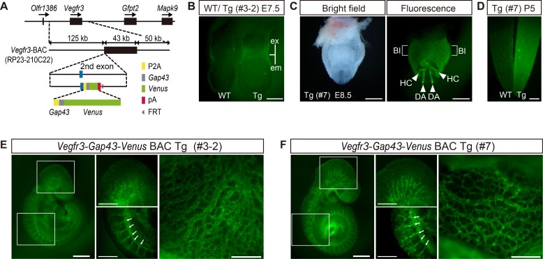
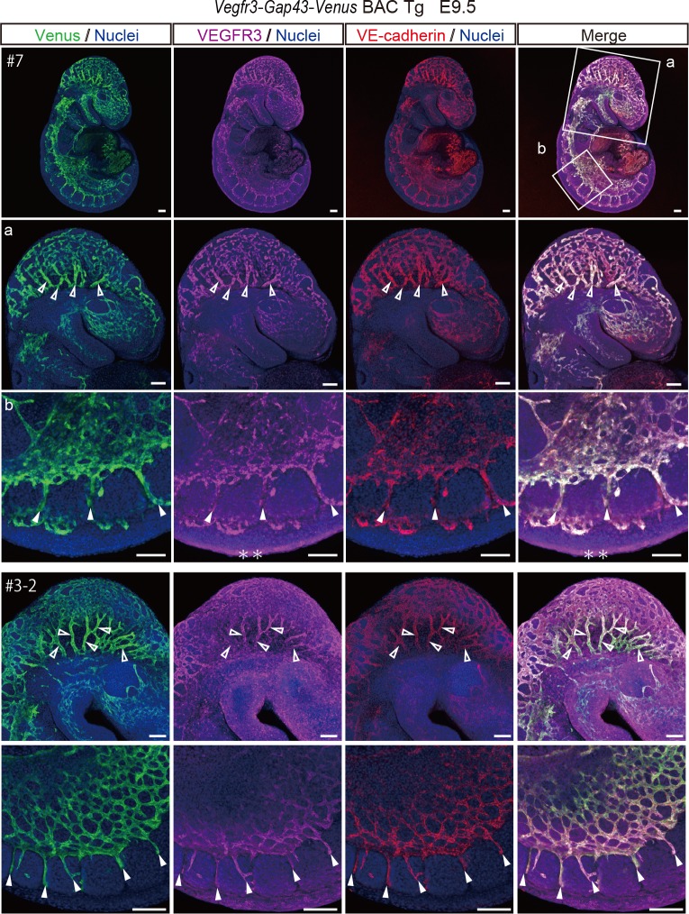
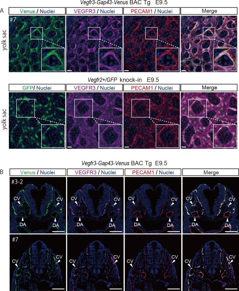
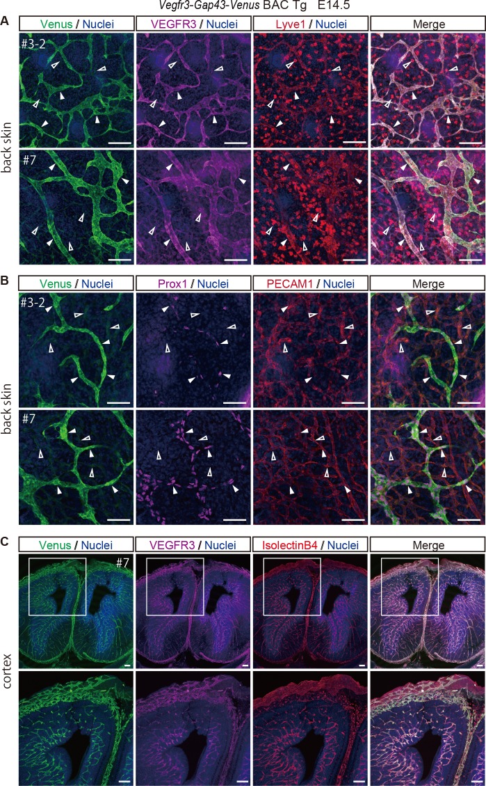
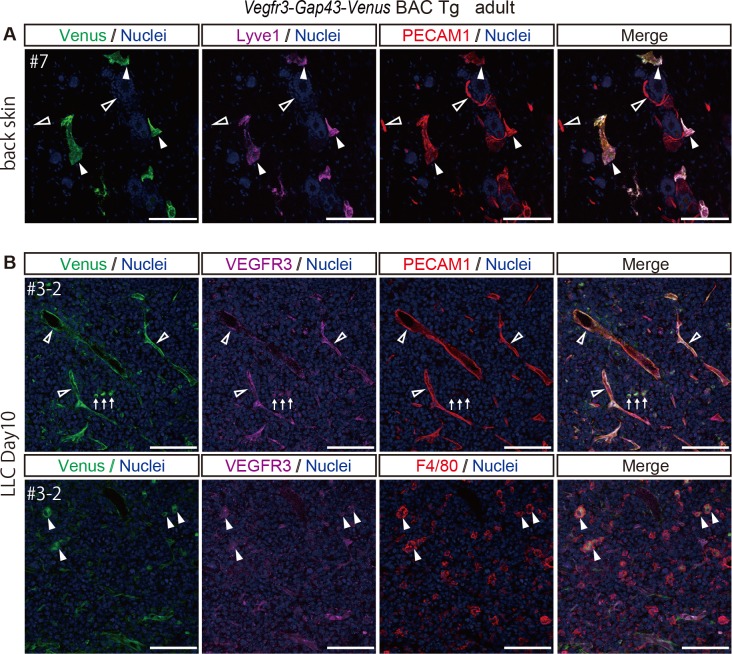
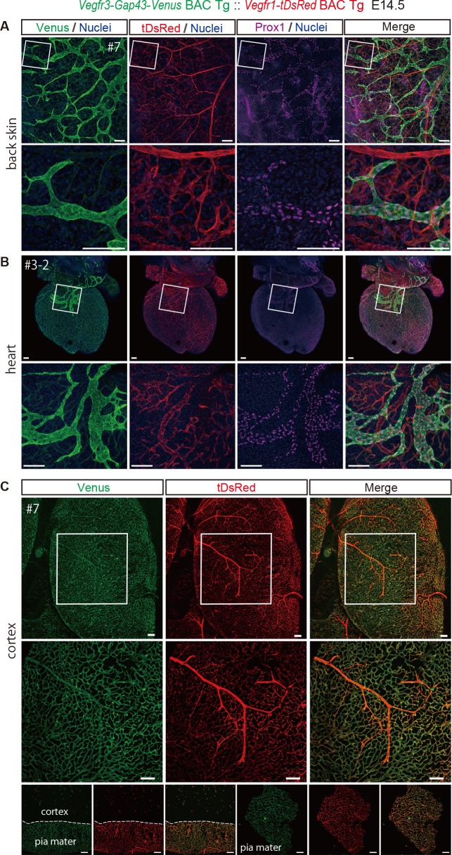
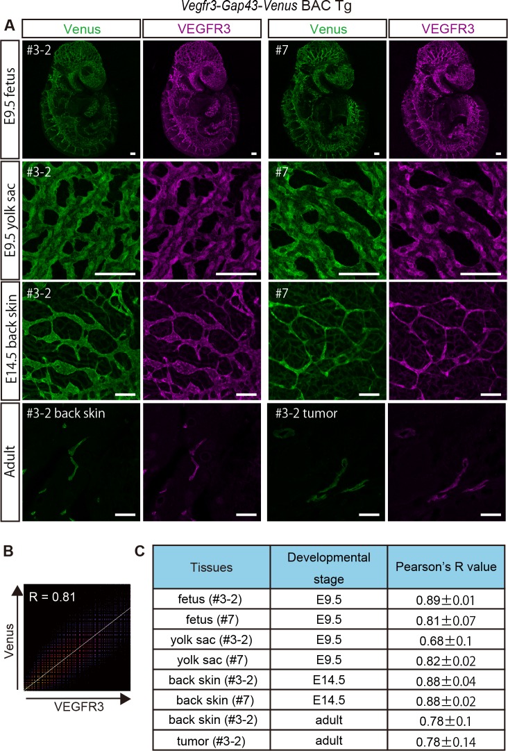
Vascular endothelial growth factor receptor 3 (Vegfr3) has been widely used as a marker for lymphatic and vascular endothelial cells during mouse embryonic development and in adult mouse, making it valuable for studying angiogenesis and lymphangiogenesis under normal and pathological conditions. Here, we report the generation of a novel transgenic (Tg) mouse that expresses a membrane-localized fluorescent reporter protein, Gap43-Venus, under the control of the Vegfr3 regulatory sequence. Vegfr3-Gap43-Venus BAC Tg recapitulated endogenous Vegfr3 expression in vascular and lymphatic endothelial cells during embryonic development and tumor development. Thus, this Tg mouse line contributes a valuable model to study angiogenesis and lymphangiogenesis in physiological and pathological contexts.


Conflict of interest statement

The authors have declared that no competing interests exist.

Figures

Fig 1
Fig 1. Generating Vegfr3-Gap43-Venus BAC Tg mice.
(A) Constructing the Vegfr3-Gap43-Venus BAC Tg. The Vegfr3 BAC clone (RP23-210C22) covering 125 kb upstream and 50 kb downstream of the Vegfr3 gene was used. Note that the PGK-gb2-neo cassette was removed from the BAC construct prior to generating Tg mice. P2A: Pocine teschovirus self-cleaving peptide sequence, Gap43: mouse Gap43 membrane localization sequence; pA: polyadenylation site. FRT: a recognition sequence for FLPe. (B) Venus expression in WT and Tg (line #3–2) embryos at E7.5. (C) Venus expression in Tg (line #7) embryos at E8.5. (D) Venus expression in WT and Tg (line #7) tails at P5. (E, F) Venus expression in Tg (line #3–2 and #7) embryos at E9.5. Arrows indicate intersomitic vessels. ex, extraembryonic; em, embryonic; Bl, blood island; HC, heart crescent; DA, dorsal aorta. Scale bar: 100 μm (B), 500 μm (C, D, E, F). All images were captured by a Leica FLIII microscope using GFP LP filter.
Fig 2
Fig 2. Venus expression in a Vegfr3-Gap43-Venus BAC Tg embryo at E9.5.
Immunofluorescence analysis of a Tg embryo at E9.5 for a vascular endothelial marker, VE-cadherin (red), Venus (anti-GFP, green), VEGFR3 (magenta) and Nuclei (Hoechst33342, blue). A higher magnification of optical sections is shown in panels a and b. Note that Venus and VEGFR3 were co-expressed in the VE-cadherin–positive endothelial cells of the Tg embryo. Open and closed arrowheads indicate vascular endothelial cells in the head region and in intersomitic vessels, respectively. Scale bar: 100 μm. * indicates non-specific signal. Images were captured by a Leica TCS-SP8 confocal microscope using a 5x/0.1 dry objective lens (Upper panel), 10x/ 0.3 dry objective lens (Middle panel) and a 20x/0.7 dry objective lens (Lower panel). Immunofluorescence analysis of the Tg embryo (line# 3–2) at E9.5 for a vascular endothelial marker, VE-cadherin (red), Venus (anti-GFP, green), VEGFR3 (magenta) and Nuclei (Hoechst33342, blue). Note that Venus and VEGFR3 are co-expressed in the VE-cadherin-positive vascular endothelial cells of the Tg embryo. Open and closed arrowheads indicate endothelial cells in the head region and intersomitic vessels, respectively. Scale bar: 100 μm. Images were captured by a Leica TCS-SP8 confocal microscope using a 10x/0.15 dry objective lens (Upper panels) and 20x/0.3 dry objective lens (Lower panels).
Fig 3
Fig 3. Membrane localization of Venus in a Vegfr3-Gap43-Venus BAC Tg yolk sac at E9.5 and Venus expression in a Vegfr3-Gap43-Venus BAC Tg embryo at E9.5.
(A) Immunofluorescence analysis of the Vegfr3-Gap43-Venus BAC Tg yolk sac at E9.5 for a vascular endothelial marker, PECAM1 (red), Venus (anti-GFP, green), VEGFR3 (magenta) and Nuclei (Hoechst33342, blue). Note that the Venus of the Vegfr3-Gap43-Venus BAC Tg yolk sac was localized in the plasma membrane of endothelial cells, while the GFP of Vegfr2+/GFP knock-in was localized in the cytoplasm. Scale bar: 20 μm. All images were captured by a Leica TCS-SP8 confocal microscope using a 40x/1.25 oil objective lens. (B) Immunofluorescence images of the Tg embryo at E9.5 for PECAM1 (red), Venus (anti-GFP, green), VEGFR3 (magenta) and Nuclei (Hoechst33342, blue). Cryosections were prepared from the Tg embryos and subjected to immunohistochemistry. Note that endogenous VEGFR3 and Venus were overlapped in the vascular endothelial cells of the Tg embryo. DA: dorsal aorta; CV: cardinal vein. Scale bar: 100 μm. All images were captured by a Leica TCS-SP8 confocal microscope using a 20x/0.7 dry objective lens.
Fig 4
Fig 4. Venus expression in a Vegfr3-Gap43-Venus BAC Tg embryo’s skin and cortex at E14.5.
(A) Immunofluorescence images of the back skin of a Tg embryo at E14.5 for Venus (anti-GFP, green), VEGFR3 (magenta), Lyve1 (red) and Nuclei (Hoechst33342, blue). Note that endogenous VEGFR3 and Venus were overlapped in the Lyve1-positive lymphatic endothelial cells of the Tg embryo. Closed arrowheads indicate Lyve1-positive lymphatic endothelial cells. Open arrowheads indicate Lyve1-positive macrophages. Scale bar: 100 μm. Images were captured by a Leica TCS-SP8 confocal microscope using a 20x/0.7 dry objective lens. (B) Immunofluorescence images of the back skin of a Tg embryo at E14.5 for Venus (anti-GFP, green), Prox1 (magenta), PECAM1 (red) and Nuclei (Hoechst33342, blue). Note that Venus was overlapped with the Prox1-positive lymphatic endothelial cells of the Tg embryo. Closed arrowheads indicate Prox1-positive lymphatic endothelial cells. Open arrowheads indicate PECAM1-positive and Prox1-negative vascular endothelial cells. Scale bar: 50 μm. All images were captured by a Leica TCS-SP8 confocal microscope using a 40x/1.25 oil objective lens. (C)Immunofluorescence images of the cortex of a Tg embryo at E14.5 for Venus (anti-GFP, green), VEGFR3 (magenta), IsolectinB4 (red) and Nuclei (Hoechst33342, blue). Note that endogenous VEGFR3 and Venus were overlapped in the IsolectinB4-stained vascular endothelial cells of the Tg embryo. Scale bar: 100 μm. Images were captured by a Leica TCS-SP8 confocal microscope using a 5x/0.15 dry objective lens (upper panels) and 10x/0.3 dry objective lens (lower panels).
Fig 5
Fig 5. Venus expression in the normal back skin and tumor of Vegfr3-Gap43-Venus BAC Tg adult mice.
(A) Immunofluorescence images of the back skin of an adult Tg mouse for Venus (anti-GFP, green), Lyve1 (magenta), PECAM1 (red) and Nuclei (Hoechst33342, blue). Closed and open arrowheads indicate Lyve1-positive lymphatic endothelial cells and Lyve1-negative vascular endothelial cells, respectively. (B) Immunofluorescence images of the tumor implanted in the Tg for Venus (anti-GFP, green), VEGFR3 (magenta), PECAM1 (upper panel) or F4/80 (lower panel) (red) and Nuclei (Hoechst33342, blue). Open arrowheads indicate PECAM1-positive vascular endothelial cells. Arrows indicate PECAM1-negative cells with a round morphology. Closed arrowheads indicate F4/80-positive macrophages. Scale bar: 100 μm. All images were captured by a Leica TCS-SP8 confocal microscope using a 20x/0.7 dry objective lens.
Fig 6
Fig 6. Simultaneous detection of Vegfr3-Gap43-Venus and Vegfr1-tDsRed in the skin, heart and brain cortex at E14.5.
(A) Immunofluorescence images of the back skin of Tg (#7) at E14.5 embryos for Venus, tDsRed and Prox1. Note that Venus expression is overlapped with that of Prox1, but not tDsRed in vascular endothelial cells. Scale bar: 100 μm. Images were captured by a Leica TCS-SP8 confocal microscope using a 10x/0.3 dry objective lens (Upper panels) and a 40x/1.25 oil objective lens (Lower panels). (B) Frontal view of immunofluorescence images of the heart of a Tg (#3–2) mouse at E14.5 for Venus, tDsRed and Prox1. Images were captured by a Leica TCS-SP8 confocal microscope using a 5x/0.15 dry objective lens (A, B Upper panels), 10x/0.3 dry objective lens (A Lower panels), and 20x/0.7 dry objective lens (B Lower panels). (C) Top view of the brain cortex of Tg (#7) at E14.5 for Venus and tDsRed. An unstained sample treated with 2%PFA was processed for imaging. Bottom panels indicate direct fluorescence of pia mater and cortex. Scale bar: 100 μm.
Fig 7
Fig 7. Quantitative colocalization analysis of endogenous VEGFR3 and Venus in Vegfr3-Gap43-Venus BAC Tg at various developmental stages.
(A) Expression of endogenous VEGFR3 and Venus in Tg (#3–2, #7) at various developmental stages. (B) A 2D intensity histogram for Pearson’s R between VEGFR3 and Venus. (C) Summary of Pearson’s R between VEGFR3 and Venus at various developmental stages.
Fig 8
Fig 8. Detection of pERK signaling of vascular endothelial cells in Vegfr3-Gap43-Venus Tg embryos at E9.5.
(A) Flow-cytometry analysis of Vegfr3-Gap43-Venus Tg embryos at E9.5. (B) Western blot analysis of Venus-positive and -negative cells from Tg embryos. (C) Phosphorylated ERK signaling in Venus-positive cells cultured in the presence or absence of the MEK inhibitor. (D) Percentage of pERK-positive cells in Venus-positive cells.

References

    1. Shibuya M, Claesson-Welsh L. Signal transduction by VEGF receptors in regulation of angiogenesis and lymphangiogenesis. Exp Cell Res. 2006;312: 549–60. 10.1016/j.yexcr.2005.11.012 - DOI - PubMed
    1. Matsumoto K, Ema M. Roles of VEGF-A signalling in development, regeneration, and tumours. J Biochem. 2014;156: 1–10. 10.1093/jb/mvu031 - DOI - PubMed
    1. Deng Y, Zhang X, Simons M. Molecular controls of lymphatic VEGFR3 signaling. Arterioscler Thromb Vasc Biol. 2015;35: 421–9. 10.1161/ATVBAHA.114.304881 - DOI - PMC - PubMed
    1. Ferrara N, Gerber H-P, LeCouter J. The biology of VEGF and its receptors. Nat Med. 2003;9: 669–76. 10.1038/nm0603-669 - DOI - PubMed
    1. Kaipainen A, Korhonen J, Mustonen T, van Hinsbergh VW, Fang GH, Dumont D, et al. Expression of the fms-like tyrosine kinase 4 gene becomes restricted to lymphatic endothelium during development. Proc Natl Acad Sci U S A. 1995;92: 3566–70. Available: http://www.ncbi.nlm.nih.gov/pubmed/7724599 - PMC - PubMed

Publication types

MeSH terms

Substances