

Circumscribed choroidal haemangioma: clinical and topographical features
- PMID: 30602446
- PMCID: PMC6817701
- DOI: 10.1136/bjophthalmol-2018-313388
Circumscribed choroidal haemangioma: clinical and topographical features
Abstract
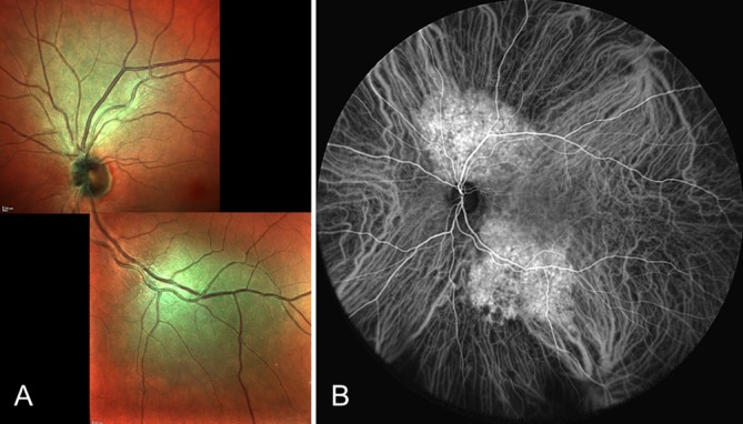
Aims: To characterise the clinical and topographical features of circumscribed choroidal haemangioma (CCH) and to visualise the patterns of tumour extent in the ocular fundus.
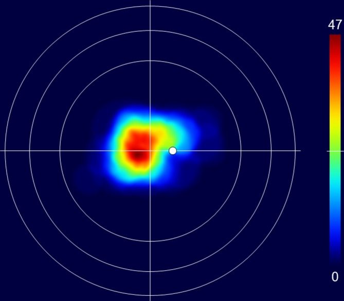
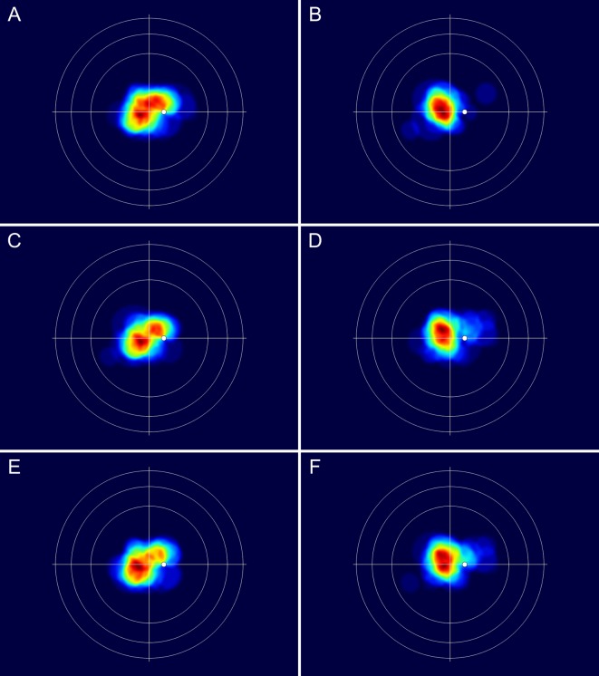
Methods: Data on the size, shape and location of 113 CCH were converted into a database of two-dimensional retinal charts by means of computer drawing software. The extent of the tumours was visualised by merging the charts and displaying the number of overlapping tumours on colour-coded maps.
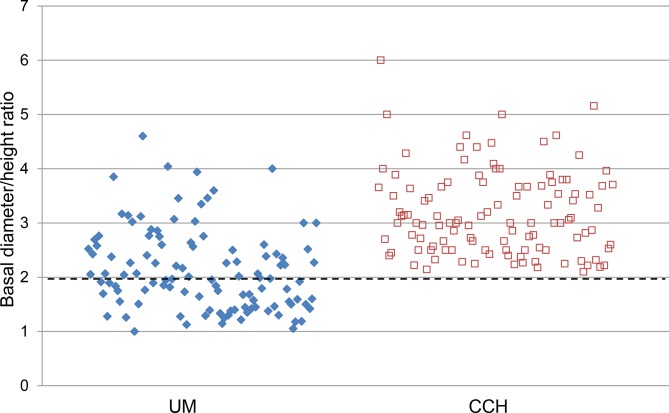
Results: The mean largest tumour diameter was 7.2 mm (range, 2.5-11.0 mm), mean tumour height was 2.4 mm (range, 0.7-4.6 mm) and mean diameter/height ratio was 3.2 (range, 2.1-6.0). The mean distance from the posterior tumour margin to the foveola and optic disc margin was 1.7 mm (range, 0-15 mm) and 2.4 mm (range, 0-11 mm), respectively. The hemispheric location of the tumour centroid was temporal in 75 eyes (66%) and nasal in 38 (34%) (p=0.0005) and the distribution between the superior and inferior hemispheres was 68 (60%) and 45 (40%), respectively (p=0.03). The presence of subretinal fluid (SRF) was significantly associated with young age at diagnosis (p=0.0002), low tumour diameter/height ratio (p=0.0004), nasal hemisphere location (p=0.006) and close proximity to the optic disc (p=0.004).
Conclusions: The superotemporal quadrant close to the macula is the most frequent location of CCH. The tumours are generally characterised by a diameter/height ratio of >2. Tumours in young patients, with marked elevation, in nasal hemisphere and in proximity to the optic disc are associated with SRF exudation.
Keywords: circumscribed choroidal haemangioma; distribution; imaging; location; topography.
© Author(s) (or their employer(s)) 2019. Re-use permitted under CC BY-NC. No commercial re-use. See rights and permissions. Published by BMJ.
Conflict of interest statement
Competing interests: None declared.
Figures
References
-
- Singh AD, Kaiser PK. Uveal vascular tumors : Singh AD, Damato BE, Pe’er J, Clinical ophthalmic oncology. Philadelphia, PA: Saunders Elsevier, 2007: 289–99.
-
- Jarrett WH, Hagler WS, Larose JH, et al. Clinical experience with presumed hemangioma of the choroid: radioactive phosphorus uptake studies as an aid in differential diagnosis. Trans Sect Ophthalmol Am Acad Ophthalmol Otolaryngol 1976;81:862–70. - PubMed
-
- Verbeek AM, Koutentakis P, Deutman AF. Circumscribed choroidal hemangioma diagnosed by ultrasonography. A retrospective analysis of 40 cases. Int Ophthalmol 1995;19:185–9. - PubMed
Publication types
MeSH terms
Substances
LinkOut - more resources
Full Text Sources
Medical
Miscellaneous