Review
. 2019 Mar 7;14(3):454-468.
doi: 10.2215/CJN.08020718. Epub 2019 Jan 2.

MicroRNAs in AKI and Kidney Transplantation

Affiliations
Review

MicroRNAs in AKI and Kidney Transplantation

Kristien J Ledeganck et al. Clin J Am Soc Nephrol. .

Abstract

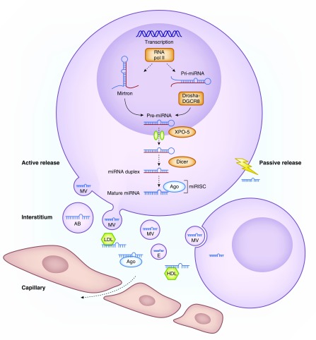
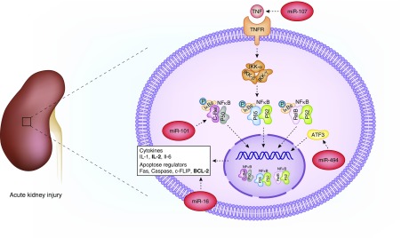
MicroRNAs are epigenetic regulators of gene expression at the posttranscriptional level. They are involved in intercellular communication and crosstalk between different organs. As key regulators of homeostasis, their dysregulation underlies several morbidities including kidney disease. Moreover, their remarkable stability in plasma and urine makes them attractive biomarkers. Beyond biomarker studies, clinical microRNA research in nephrology in recent decades has focused on the discovery of specific microRNA signatures and the identification of novel targets for therapy and/or disease prevention. However, much of this research has produced equivocal results and there is a need for standardization and confirmation in prospective trials. This review aims to provide an overview of general concepts and available clinical evidence in both the pathophysiology and biomarker fields for the role of microRNA in AKI and kidney transplantation.

Keywords: Acute Kidney Injury; Biomarkers; Body Fluids; Homeostasis; MicroRNAs; Prospective Studies; kidney transplantation.


Figures

Figure 1.
Figure 1.
MicroRNA biogenesis and function. microRNA coding regions in the human genome are found either intergenic or in the introns of annotated genes. microRNA synthesis starts in the nucleus where most of the miRs are transcribed by RNA polymerase II into primary miR transcripts (pri-miR) of several kilobases that contain local stem-loop structures. The first step of miR maturation is cleavage at the stem of the hairpin structure by a microprocessor complex consisting of Drosha (an RNase III protein) together with its cofactor DiGeorge Syndrome Critical Region 8 (DGCR8), which releases a small hairpin structure of 70 nucleotides that is termed a precursor miR (pre-miR). After nuclear processing, pre-miRs are exported to the cytoplasm by exportin 5 (XPO-5), where they are cleaved near the terminal loop by another RNase enzyme called Dicer, thereby releasing an approximately 22 nucleotide miR duplex. This duplex is loaded onto an AGO protein to generate the microRNA-induced silencing complex (miRISC). One strand (guide strand) remains in the AGO protein as a biologically active miR whereas the other stand (passenger strand, known as miR*) is degraded. The mature miR as part of the effector RISC binds to the 3′UTR region of the mRNA and mediates mRNA degradation, destabilization, or translational inhibition. Apart from this canonical pathway, there is an alternative “mirtron” pathway, independent from Drosha and DGCR8. Mirtrons are miRs that originate from spliced-out introns and are created when small RNAs bind to the termini of small intronic hairpins. Pre-microRNA hairpins with 3′ overhangs are so formed and can mature into 22 nucleotides structures, which look and function as normal miRs. microRNAs exert their repressive function intracellularly, but are also released into the extracellular compartment, with this initiating their role as important intercellular communicators as they are taken up by recipient cells. microRNA can be released passively after cell death or injury, or can be actively secreted in different types of extracellular vesicles, including exosomes, microvesicles and apoptotic bodies. Circulating microRNAs form complexes with RNA binding proteins including AGO2 proteins and lipoproteins (HDL and LDL), which protects them from RNAse-dependent degradation. AB, apoptotic body; E, exosome; MV, microvesicle; RNA pol II, RNA polymerase 2.
Figure 2.
Figure 2.
Several microRNAs are involved in the NFκB pathway in AKI. Several microRNA contribute to AKI by targeting the NFκB pathway. ATF3, activating transcription factor 3; BCL-2, B cell lymphoma-2; c-FLIP, cellular FLICE-like inhibitory protein; c-Rel, P50, P52, P65 and RelB, NF-κB transcription factor family members; Fas, first apoptosis signal; IKK, IκB kinase; TNFR, TNF receptor.
Figure 3.
Figure 3.
Several specific microRNAs are up- or downregulated in the kidney graft in transplantation-related kidney disease. Functional processes are represented in the inner circle, and the outer circle represents molecular pathways of interest for the particular clinical entity. Only the biologically confirmed microRNA–target interactions are highlighted. The color code of the microRNAs indicates whether the tissue microRNA are mimicked in biofluids. Red represents parallel findings in plasma/serum for that particular microRNA. Orange represents parallel findings in urine for that particular microRNA and blue represents parallel findings in PBMC. Combination of the colors is possible. ABMR, antibody-mediated rejection; BCL-2, B cell lymphoma-2; CDKN2, cyclin-dependent kinase inhibitor 2; CXCL8, C-X-X motive chemokine ligand 8; ECM, extracellular matrix; EDN, endothelin; IRI/DGF, ischemia/reperfusion injury and delayed graft function; NFκB/CXCL-8, NFκB/C-X-C motive chemokine ligand 8; TCMR, T cell–mediated rejection; TGFβ/FOXP3, TGFβ/forkhead box P3; TGFβ/SMAD7, TGFβ/SMAD family member 7; VEGF, vascular endothelial growth factor.

References

    1. Michell DL, Vickers KC: Lipoprotein carriers of microRNAs. Biochim Biophys Acta 1861[12 Pt B]: 2069–2074, 2016 - PMC - PubMed
    1. Bayraktar R, Van Roosbroeck K, Calin GA: Cell-to-cell communication: MicroRNAs as hormones. Mol Oncol 11: 1673–1686, 2017 - PMC - PubMed
    1. Zhang W, Zhou X, Zhang H, Yao Q, Liu Y, Dong Z: Extracellular vesicles in diagnosis and therapy of kidney diseases. Am J Physiol Renal Physiol 311: F844–F851, 2016 - PMC - PubMed
    1. Zhang A, Li M, Wang B, Klein JD, Price SR, Wang XH: miRNA-23a/27a attenuates muscle atrophy and renal fibrosis through muscle-kidney crosstalk. J Cachexia Sarcopenia Muscle 9: 755–770, 2018 - PMC - PubMed
    1. Rupaimoole R, Slack FJ: MicroRNA therapeutics: Towards a new era for the management of cancer and other diseases. Nat Rev Drug Discov 16: 203–222, 2017 - PubMed

Publication types

MeSH terms