. 2019 Mar 8;294(10):3670-3682.
doi: 10.1074/jbc.RA118.004671. Epub 2019 Jan 2.

p53 reactivation with induction of massive apoptosis-1 (PRIMA-1) inhibits amyloid aggregation of mutant p53 in cancer cells

Affiliations

p53 reactivation with induction of massive apoptosis-1 (PRIMA-1) inhibits amyloid aggregation of mutant p53 in cancer cells

Luciana P Rangel et al. J Biol Chem. .

Abstract

p53 mutants can form amyloid-like structures that accumulate in cells. p53 reactivation with induction of massive apoptosis-1 (PRIMA-1) and its primary active metabolite, 2-methylene-3-quinuclidinone (MQ), can restore unfolded p53 mutants to a native conformation that induces apoptosis and activates several p53 target genes. However, whether PRIMA-1 can clear p53 aggregates is unclear. In this study, we investigated whether PRIMA-1 can restore aggregated mutant p53 to a native form. We observed that the p53 mutant protein is more sensitive to both PRIMA-1 and MQ aggregation inhibition than WT p53. The results of anti-amyloid oligomer antibody assays revealed that PRIMA-1 reverses mutant p53 aggregate accumulation in cancer cells. Size-exclusion chromatography of the lysates from mutant p53-containing breast cancer and ovarian cell lines confirmed that PRIMA-1 substantially decreases p53 aggregates. We also show that MDA-MB-231 cell lysates can "seed" aggregation of the central core domain of recombinant WT p53, corroborating the prion-like behavior of mutant p53. We also noted that this aggregation effect was inhibited by MQ and PRIMA-1. This study provides the first demonstration that PRIMA-1 can rescue amyloid-state p53 mutants, a strategy that could be further explored as a cancer treatment.

Keywords: amyloid; anticancer drug; p53; protein aggregation; protein misfolding.


Conflict of interest statement

The authors declare that they have no conflicts of interest with the contents of this article

Figures

Figure 1.
Figure 1.
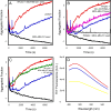
PRIMA-1 and MQ inhibit both R248Qp53C and WTp53C aggregation at 37 °C. The samples (5 μm) were incubated at 37 °C for 1 h in the presence of PRIMA-1, MQ, or 0.1% DMSO (final concentration) as the control. A, WTp53C aggregation in the absence (black) or presence of 100 μm PRIMA-1 (green line) or 100 μm MQ (red line). B, R248Qp53C aggregation in the absence (black line) or presence of 100 μm PRIMA-1 (red) or 100 μm MQ (green line). C, lower concentrations of MQ inhibited R248Qp53C aggregation: control R248Qp53C (dark red line), 10 μm MQ (dark yellow line), 25 μm MQ (orange line), 50 μm (green line), and 100 μm MQ (blue line). D, MQ dose-dependent inhibition of R248Q mutant p53 aggregation at 37 °C after 2 h. Protein aggregation was assessed by monitoring the increase in the ThT fluorescence intensity (excitation, 440 nm; emission, 482 nm) at a ThT:protein ratio of 5:1. All samples were diluted in 50 mm Tris (pH 7.2), 150 mm NaCl, 5 mm DTT, and 5% glycerol. This figure shows one representative experiment; n = 4.
Figure 2.
Figure 2.
Human p73 central core domain (p73C) aggregation in the presence of MQ. MQ shows a small effect on p73C aggregation after 4 h at 37 °C. A, p73C at 5 μm was submitted to aggregation at 37 °C for 4 h, monitored by ThT binding at 25 μm. Black line, p73C; red line, p73C treated with 100 μm MQ. B, p73C light scattering (LS), before (soluble p73C and soluble p73C + 100 μm MQ) and after the 4-h kinetics (aggregated p73C and aggregated p73C + 100 μm MQ). All samples were diluted in 50 mm Tris (pH 7.2), 150 mm NaCl, 5 mm DTT, and 5% glycerol. This figure shows one representative experiment; n = 2.
Figure 3.
Figure 3.
Mutant p53 and amyloid oligomer staining are reduced in the MDA-MB-231 cell line after PRIMA-1 treatment in a concentration-dependent manner. A, MDA-MB-231 cells, treated with 25, 50, or 100 μm PRIMA-1 or 0.1% DMSO as the control for 16 h, were labeled with anti-p53 (DO-1) and anti-oligomer (A11) antibodies. Nuclei were stained with DAPI. Columns from left to right show nuclear staining with DAPI, p53 labeling, labeling of amyloid oligomers, and merged images. White arrowheads indicate cells stained with either DO-1 or A11 antibodies in their correspondent panels. Magnification, ×400. Scale bar = 50 μm.
Figure 4.
Figure 4.
A, MCF-7 cells were treated for 16 h with either 100 μm PRIMA-1 or 0. 1% DMSO as a control. The cells were labeled with anti-p53 (DO-1) and anti-oligomer (A11) antibodies, and nuclei were stained with DAPI. Columns from left to right show DAPI, p53 labeling, labeling of amyloid oligomers, and merged images. Magnification, ×400. Scale bar = 50 μm. B, MTT assay showing MDA-MB-231 (black line) and MCF-7 (red line) cell viability after treatment for 24 h with a serial dilution of PRIMA-1 from 0 (0.2% DMSO) to 200 μm. n = 4. C, cell proliferation assay using trypan blue showing a dose-dependent inhibition of cell proliferation after 24 h of treatment with PRIMA-1. n = 3 and significance marked with *(p < 0.05) and with *** (p < 0.001). D, AnnexinV/PI assay showing that PRIMA-1 induces apoptosis of MDA-MB-231 cells at 50 μm PRIMA-1, showing its effectiveness in this cell line in the concentration range used in this study.
Figure 5.
Figure 5.
Dot blot of cancer cell lysates showing that the amyloid oligomer levels are reduced by PRIMA-1 treatment. A, dot blots for A11 were performed with equal protein amounts from lysates of MDA-MB-231, MCF-7, or OVCAR-3 cells treated with 100 μm PRIMA-1 or 0.1% DMSO. Aggregated R248Qp53C at 20 μm and transthyretin (TTR) at 10 μm were used as positive controls, and soluble R248Qp53C at 20 μm and 0.1% BSA were used as negative controls. B, quantification of dot blots from a total of four independent experiments. A significant reduction in amyloid oligomers levels in cells treated with PRIMA-1 is indicated by asterisks (***, p < 0.001; *, p < 0.05). Quantification was performed using ImageJ software (version 1.43r, National Institutes of Health). C, ThT fluorescence in the presence of lysates from cells with different p53 statuses. ThT spectra were obtained after incubation of 15 μg of protein from different cell lysates with 25 μm ThT. The result is representative of three independent measurements.
Figure 6.
Figure 6.
The p53 amyloid fraction of mutant p53 cancer cells is mobilized by PRIMA-1 treatment. A, Western blotting of lysates of MDA-MB-231 control cells treated with 0.1% DMSO or 100 μm PRIMA-1 for 18 h and quantification of p53 and MDM2 levels. n = 5. β-Actin was used as the reference. B, MDA-MB-231 cell lysates treated with PRIMA-1 or DMSO were immunoprecipitated with DO-1 and A11 and immunoblotted for p53 (anti-p53 rabbit polyclonal antibody). n = 3. C, detection and quantification of MDM2 co-immunoprecipitation. n = 3. D, immunoprecipitation assays using MCF-7 cells and their quantification. n = 3. E, immunoprecipitation assay using OVCAR-3 cells and their quantification. n = 3. *, p < 0.05. Overall, mouse IgG was used as an antibody control.
Figure 7.
Figure 7.
Size-exclusion chromatography of cell lysates. A and B, MDA-MB-231 (A) and OVCAR3 (B) cell lysates were subjected to size-exclusion chromatography for detection of p53 aggregates in cells treated with either 100 μm PRIMA-1 (red line) or DMSO (black line). Aggregates eluted in the column void volume. Fractions eluted were separated through SDS-PAGE and immunoblotted for p53 (DO1). n = 3.
Figure 8.
Figure 8.
Seeding of WT p53 aggregation by MDA-MB-231 protein extract. A–C, WTp53C at 5 μm (blue lines), protein extract at 3 μg/ml incubated with WTp53C at 5 μm (red lines), and control of protein extract alone used as the seed at 3 μg/ml (black lines). B, MDA-MB-231 cell extract treated with 3 μg/ml PRIMA-1 was incubated with 5 μm WTp53C (magenta line). MDA-MB-231 cell extract treated with PRIMA-1 was used as the seed at 3 μg/ml (gray line). The traces for WTp53C (blue line), protein extract incubated with WTp53C (red line), and control of protein extract alone used as the seed (black line) from A are repeated in the panel. C, MDA-MB-231 protein extract at 3 μg/ml was incubated with 5 μm WTp53C and 100 μm MQ (green line); the traces for WTp53C (blue line), protein extract incubated with WTp53C (red line), and control of protein extract alone used as the seed (black line) from A are repeated in the panel. D, ThT spectra (excitation, 450 nm; emission, 480 to 550 nm) after the 2-h kinetics experiment in A: WTp53C at 5 mm (blue line), protein extract at 3 μg/ml incubated with WTp53C at 5 μm (red line), and control of protein extract alone used as the seed at 3 μg/ml (yellow line). Aggregation was monitored by thioflavin T fluorescence emission (excitation, 440 nm; emission, 482 nm) over time at 37 °C. The ThT concentration was 25 μm, and measurements were performed at pH 7.2 in 50 mm Tris buffer, 150 mm NaCl, 5% glycerol, and 5 mm DTT. Aggregated fraction = (Fobs − FI)/(FF − FI), where F is the ThT fluorescence emission, Fobs is the observed fluorescence emission, FI is the initial fluorescence, and FF is the final fluorescence. The figure shows an experiment representative of three independent experiments.
Figure 9.
Figure 9.
Summary of PRIMA-1/MQ effects on amyloid-state p53. 1, PRIMA-1 is converted to MQ in biological systems, as described by Lambert et al. (29). 2, PRIMA-1 and MQ inhibit p53C aggregation in vitro. 3, PRIMA-1 mobilizes the aggregated p53 pool and stabilizes mutant p53, resulting in recovery of WT p53 functions and a reduction in cellular amyloid content. 4, PRIMA-1/MQ blocks the seeding of WT p53C aggregation by mutant p53 present in cell lysates, which might be part of the general effect of PRIMA-1/MQ on p53 seen in mutant p53 cancer cells.

Similar articles

Cited by

References

    1. Freed-Pastor W. A., and Prives C. (2012) Mutant p53: One name, many proteins. Genes Dev. 26, 1268–1286 10.1101/gad.190678.112 - DOI - PMC - PubMed
    1. Vousden K. H., and Lane D. P. (2007) p53 in health and disease. Nat. Rev. Mol. Cell Biol. 8, 275–283 10.1038/nrm2147 - DOI - PubMed
    1. Alexandrova E. M., Yallowitz A. R., Li D., Xu S., Schulz R., Proia D. A., Lozano G., Dobbelstein M., and Moll U. M. (2015) Improving survival by exploiting tumour dependence on stabilized mutant p53 for treatment. Nature 523, 352–356 10.1038/nature14430 - DOI - PMC - PubMed
    1. Zhu J., Sammons M. A., Donahue G., Dou Z., Vedadi M., Getlik M., Barsyte-Lovejoy D., Al-awar R., Katona B. W., Shilatifard A., Huang J., Hua X., Arrowsmith C. H., and Berger S. L. (2015) Gain-of-function p53 mutants co-opt chromatin pathways to drive cancer growth. Nature 525, 206–211 10.1038/nature15251 - DOI - PMC - PubMed
    1. Muller P. A., and Vousden K. H. (2014) Mutant p53 in cancer: new functions and therapeutic opportunities. Cancer Cell 25, 304–317 10.1016/j.ccr.2014.01.021 - DOI - PMC - PubMed

Publication types