. 2019 Mar 5;93(6):e02066-18.
doi: 10.1128/JVI.02066-18. Print 2019 Mar 15.

Inhibition of Ongoing Influenza A Virus Replication Reveals Different Mechanisms of RIG-I Activation

Affiliations

Inhibition of Ongoing Influenza A Virus Replication Reveals Different Mechanisms of RIG-I Activation

GuanQun Liu et al. J Virol. .

Abstract

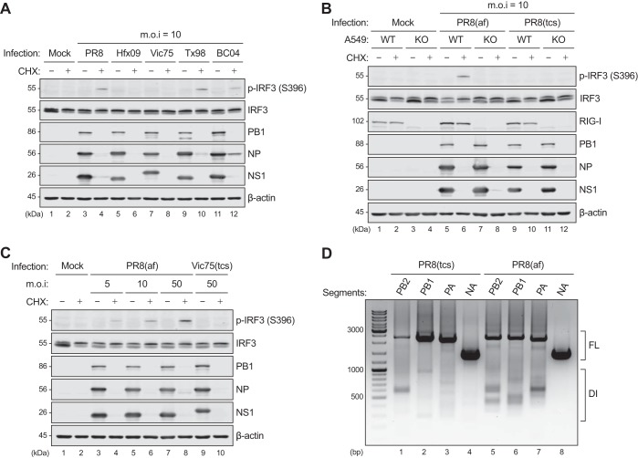
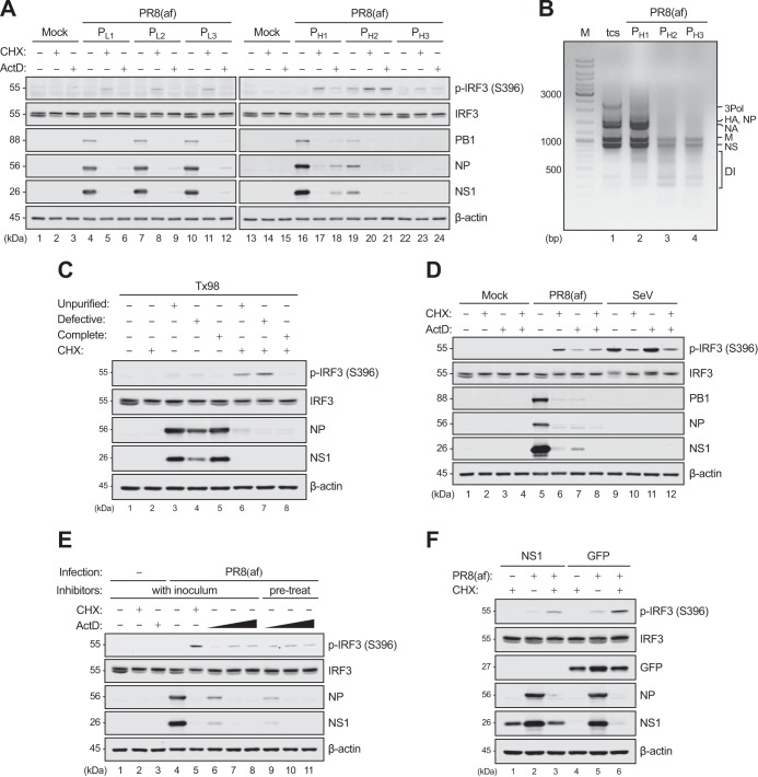
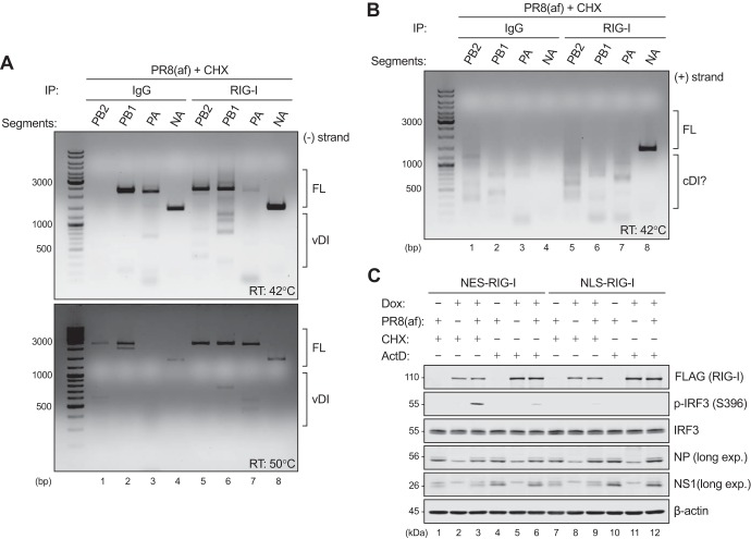
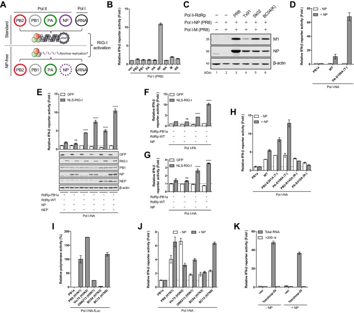
Pattern recognition receptors provide essential nonself immune surveillance within distinct cellular compartments. Retinoic acid-inducible gene I (RIG-I) is one of the primary cytosolic RNA sensors, with an emerging role in the nucleus. It is involved in the spatiotemporal sensing of influenza A virus (IAV) replication, leading to the induction of type I interferons (IFNs). Nonetheless, the physiological viral ligands activating RIG-I during IAV infection remain underexplored. Other than full-length viral genomes, cellular constraints that impede ongoing viral replication likely potentiate an erroneous viral polymerase generating aberrant viral RNA species with RIG-I-activating potential. Here, we investigate the origins of RIG-I-activating viral RNA under two such constraints. Using chemical inhibitors that inhibit continuous viral protein synthesis, we identify the incoming, but not de novo-synthesized, viral defective interfering (DI) genomes contributing to RIG-I activation. In comparison, deprivation of viral nucleoprotein (NP), the key RNA chain elongation factor for the viral polymerase, leads to the production of aberrant viral RNA species activating RIG-I; however, their nature is likely to be distinct from that of DI RNA. Moreover, RIG-I activation in response to NP deprivation is not adversely affected by expression of the nuclear export protein (NEP), which diminishes the generation of a major subset of aberrant viral RNA but facilitates the accumulation of small viral RNA (svRNA). Overall, our results indicate the existence of fundamentally different mechanisms of RIG-I activation under cellular constraints that impede ongoing IAV replication.IMPORTANCE The induction of an IFN response by IAV is mainly mediated by the RNA sensor RIG-I. The physiological RIG-I ligands produced during IAV infection are not fully elucidated. Cellular constraints leading to the inhibition of ongoing viral replication likely potentiate an erroneous viral polymerase producing aberrant viral RNA species activating RIG-I. Here, we demonstrate that RIG-I activation during chemical inhibition of continuous viral protein synthesis is attributable to the incoming DI genomes. Erroneous viral replication driven by NP deprivation promotes the generation of RIG-I-activating aberrant viral RNA, but their nature is likely to be distinct from that of DI RNA. Our results thus reveal distinct mechanisms of RIG-I activation by IAV under cellular constraints impeding ongoing viral replication. A better understanding of RIG-I sensing of IAV infection provides insight into the development of novel interventions to combat influenza virus infection.

Keywords: Influenza A virus; RIG-I; aberrant RNA; defective interfering RNA; innate immunity; interferon.


Figures

FIG 1
FIG 1
CHX unmasking of RIG-I activation by IAV strains propagated in embryonated chicken eggs, but not in tissue culture. (A) A549 cells were infected with a panel of IAV strains (PR8-H1N1, Hfx09-H1N1pdm, Vic75-H3N2, Tx98-swH3N2, and BC04-chH7N3) at an MOI of 10 in the absence or presence of CHX (50 μg/ml) for 8 h. (B) WT or RIG-I KO A549 cells were infected with PR8 stocks propagated in embryonated chicken eggs (af) or MDCK cells (tcs) (MOI =10) in the absence or presence of CHX for 8 h. (C) A549 cells were infected with increasing doses of allantoic fluid stock of PR8 (MOI =5, 10, and 50) or tissue culture stock of Vic75 (MOI = 50) in the absence or presence of CHX for 8 h. The cell lysates were subjected to immunoblotting for phosphorylated (p) IRF3 (S396), total IRF3, RIG-I, PB1, NP, NS1, and β-actin. (D) Total RNA extracted from the tissue culture (tcs) or allantoic fluid (af) stock of PR8 was subjected to S-RT-PCR to amplify the DI RNA species derived from the three viral polymerase genes. Amplification of the NA segment served as a negative control for DI presence, since it is not a major source of DI RNA generation. FL, full length.
FIG 2
FIG 2
Allantoic-fluid-derived DI genomes activate RIG-I in the presence of CHX or ActD. (A) The allantoic fluid (af) stock of PR8 was consecutively passaged in MDCK cells at a low or high MOI to obtain the PL1 to PL3 and PH1 to PH3 stocks, respectively. A549 cells were subsequently infected with these stocks (equal volumes) in the absence or presence of CHX (50 μg/ml) or ActD (1 μg/ml) for 8 h. The protein expression levels were determined by immunoblotting for phosphorylated IRF3 (S396), total IRF3, PB1, NP, NS1, and β-actin. (B) Equal amounts of total RNA extracted from the PH1 to PH3 stocks were subjected to M-RT-PCR to detect the DI RNA contents. The tissue culture supernatant (tcs) stock of PR8 served as a control with low DI content. (C) The allantoic fluid stock of Tx98 (H3N2) was purified through a sucrose gradient, and the band corresponding to the defective or complete viral particles was collected. A549 cells were infected in the absence or presence of CHX with the unpurified, defective, or complete virus at equal HA titers (128 hemagglutinating units [HAU]/ml) for 8 h. (D) A549 cells were infected with the allantoic fluid stock of PR8 (MOI = 50) or SeV-Cantell (50 HAU/ml) in the presence of CHX or ActD alone, or CHX and ActD in combination, for 8 h. (E) Increasing doses of ActD (0.2, 1, and 5 μg/ml) were added to A549 cells 1 h prior to PR8(af) inoculation (pretreatment) or along with the virus inoculum. The cells were further infected in the presence of inhibitors for 8 h. (F) A549 cells were transfected with a plasmid expressing PR8-NS1 or GFP for 24 h, followed by PR8(af) infection (MOI = 50) in the absence or presence of CHX for 8 h. (C to F) Protein expression levels were determined by immunoblotting as for panel A.
FIG 3
FIG 3
Allantoic-fluid-derived DI genomes associate with and activate cytoplasmic RIG-I. (A) A549 cells were infected with the allantoic fluid (af) stock of PR8 (MOI = 50) in the presence of CHX (50 μg/ml) for 6 h. RIP was performed in the whole-cell lysates using either the RIG-I antibody or an IgG isotype control. The associated RNAs were extracted from the respective immunoprecipitates and subjected to S-RT-PCR to amplify the DI species of negative (−) polarity (vDI) from the three polymerase and NA genes. The RT step was performed at 42°C (top) or 50°C (bottom) to increase the specificity of DI amplification. (B) The extracted RNA from the experiment shown in panel A was subjected to S-RT-PCR to amplify the DI species of positive (+) polarity (cDI) from the three polymerase and NA genes. The RT step was performed at 42°C. (C) RIG-I KO A549 cells inducibly expressing NES- or NLS-RIG-I were left noninduced or induced with 1 μg/ml doxycycline (Dox) for 4 h, followed by mock infection or infection with PR8(af) (MOI = 50) in the presence of CHX (50 μg/ml) or ActD (1 μg/ml) for 8 h. The cell lysates were subjected to immunoblotting for FLAG-RIG-I, phosphorylated IRF3 (S396), total IRF3, NP, NS1, and β-actin.
FIG 4
FIG 4
NP deprivation leads to de novo generation of RIG-I-activating viral RNA that is distinct from DI genomes. (A) Schematic diagram of the standard and NP-free RNP reconstitution systems in which differential viral RNA species activate RIG-I. (B) 293T cells were RNP reconstituted in the absence of NP with a Pol I vector or a Pol I construct generating each of the eight viral segments of PR8. Total RNA was extracted and transfected into 293T cells preexpressing an IFN-β promoter-driven firefly luciferase reporter (p125Luc) and a constitutively expressed Renilla luciferase construct (pTK-rLuc). (C) In each sample, 293T cells were cotransfected with three Pol II-driven plasmids expressing the viral polymerases (PB2, PB1, and PA) of PR8, Tx91, Sk02, or BC04(K627), along with two Pol I-driven plasmids generating the vRNAs of NP and M segments of PR8. The expression levels of NP and M1 were determined by immunoblotting at 24 h p.t. (D) 293T cells were RNP reconstituted with the PR8 NA segment (Pol I-NA) in the absence or presence of NP protein (Pol II-NP) using the catalytically inactive (PB1a), WT, or T (PA-D108A) viral polymerase. The extracted total RNA was tested for immunostimulatory activity as for panel B. (E) 293T cells were RNP reconstituted with the PR8 NA segment (Pol I-NA) using the catalytically inactive (PB1a) or WT polymerase in the presence of a plasmid expressing GFP or NLS-RIG-I. As indicated, plasmids expressing NP and NEP proteins were cotransfected along with p125Luc and pTK-rLuc. The cell lysates were subjected to immunoblotting to examine the protein expression levels of GFP, FLAG-RIG-I, PB1, NP, NEP, and β-actin. (F and G) 293T cells were RNP reconstituted with the PR8 PA (Pol I-PA) (F) or HA (Pol I-HA) (G) segment as for panel E. Statistical significance was determined by two-way ANOVA, followed by a Sidak posttest. ****, P < 0.0001; ns, not significant. (H) 293T cells were RNP reconstituted as for panel E with or without a plasmid expressing NP using the inactive (PB1a), WT, T, or R polymerase in the presence of NLS-RIG-I. (I) 293T cells were cotransfected with four Pol II-driven plasmids expressing the viral polymerases and NPs of various IAV strains, along with a Pol I-driven plasmid encoding a vRNA-like molecule in which the firefly luciferase open reading frame (ORF) was flanked by the 5′ and 3′ noncoding regions (NCRs) of the PR8 NA segment (Pol I-NA-fLuc). Reconstitution using the inactive polymerase (PB1a) of PR8 served as a negative control. (J) 293T cells were RNP reconstituted as for panel H using the polymerases of various IAV strains in the presence of NLS-RIG-I. (K) Two sets of 293T cells were RNP reconstituted in the absence or presence of NP with a Pol I vector or an artificial DI-like construct in which the 5′ and 3′ NCRs of the PR8 NP segment were linked through a UUCG tetraloop. At 24 h p.t., one set of cells was subjected to RNA extraction to obtain total RNAs containing small RNAs, while the other set was extracted for RNA species larger than ∼200 nt. These RNAs were tested for immunostimulatory activity as for panel B. Unless otherwise indicated, RLU were determined at 24 h p.t. and are expressed as fold changes relative to the Pol I vector (B and K) or the PB1a control (D and J). The data are shown as means ± SD of the results of three independent experiments performed in triplicate.
FIG 5
FIG 5
Accumulation of a major subset of aberrant viral RNAs driven by NP deprivation is sensitive to NEP expression. (A) 293T cells were RNP reconstituted using the catalytically inactive (PB1a) or WT viral polymerase with a vRNA-like construct in which the firefly luciferase ORF was flanked by the 5′ and 3′ NCRs of the PR8 NP segment (Pol I-NP-fLuc). As indicated, the plasmids expressing NP and NEP were cotransfected. At 48 h p.t., total RNA was extracted and subjected to Northern blotting to detect small RNA species containing the 5′ end of vRNA (5′ v-probe) or cRNA (5′ c-probe). The aberrant replication products under NP deprivation (lane 3) and the svRNA-like molecules generated in the presence of NEP (lane 5) are indicated by black and red arrowheads, respectively. U6 served as a loading control. The protein expression levels were determined by immunoblotting for PB1, NP, NEP, and β-actin. FL, full-length vRNA. (B) 293T cells were RNP reconstituted (+NP) using the inactive (PB1a) or WT viral polymerase with the NA-fLuc reporter construct (Pol I-NA-fLuc). As indicated, increasing doses of GFP (10 to 100 ng) or NEP were supplemented. The relative polymerase activity was determined at 24 h p.t. and is expressed as fold changes over the PB1a control. (C) 293T cells were RNP reconstituted as for panel B with the NA segment of Hfx09 (Pol I-NA) in the absence or presence of increasing doses of GFP or NEP. Total RNA was extracted at 48 h p.t. and subjected to primer extension analysis to detect the levels of vRNA, cRNA, and mRNA. 5S rRNA served as a loading control. (D) 293T cells were RNP reconstituted with the PR8 PB2 segment (Pol I-PB2) using the inactive (PB1a) or WT polymerase in the presence of GFP or NLS-RIG-I. As indicated, plasmids expressing NP and NEP were cotransfected along with p125Luc and pTK-rLuc. RLU were determined at 24 h p.t. and are expressed as fold changes relative to PB1a with GFP. The cell lysates were subjected to immunoblotting to examine the protein expression levels of GFP, FLAG-RIG-I, PB1, NP, NEP, and β-actin. Statistical significance was determined by two-way ANOVA, followed by a Sidak posttest. *, P < 0.05; ****, P < 0.0001; ns not significant. (E) Proposed model for nuclear RIG-I activation by intermolecular RNA duplexes formed from the complementarity between aberrant viral RNA or svRNA and the 3′ end of full-length vRNA (partial complementarity) or cRNA (full complementarity). The data are shown as means ± SD of the results of three independent experiments performed in triplicate.

Similar articles

Cited by

References

    1. Mogensen TH. 2009. Pathogen recognition and inflammatory signaling in innate immune defenses. Clin Microbiol Rev 22:240–273. doi:10.1128/CMR.00046-08. - DOI - PMC - PubMed
    1. Loo YM, Fornek J, Crochet N, Bajwa G, Perwitasari O, Martinez-Sobrido L, Akira S, Gill MA, Garcia-Sastre A, Katze MG, Gale M Jr. 2008. Distinct RIG-I and MDA5 signaling by RNA viruses in innate immunity. J Virol 82:335–345. doi:10.1128/JVI.01080-07. - DOI - PMC - PubMed
    1. Kato H, Takeuchi O, Sato S, Yoneyama M, Yamamoto M, Matsui K, Uematsu S, Jung A, Kawai T, Ishii KJ, Yamaguchi O, Otsu K, Tsujimura T, Koh CS, Reis e Sousa C, Matsuura Y, Fujita T, Akira S. 2006. Differential roles of MDA5 and RIG-I helicases in the recognition of RNA viruses. Nature 441:101–105. doi:10.1038/nature04734. - DOI - PubMed
    1. Loo YM, Gale M Jr. 2011. Immune signaling by RIG-I-like receptors. Immunity 34:680–692. doi:10.1016/j.immuni.2011.05.003. - DOI - PMC - PubMed
    1. Shaw ML, Palese P. 2013. Orthomyxoviridae, p 1151–1185. In Knipe DM, Howley PM (ed), Fields Virology, 6th ed, vol I Lippincott Williams & Wilkins, Philadelphia, PA, USA.

Publication types

MeSH terms