

What's Happening on the Other Side? Revealing Nano-Meter Scale Features of Mammalian Cells on Engineered Textured Tantalum Surfaces
- PMID: 30602684
- PMCID: PMC6337376
- DOI: 10.3390/ma12010114
What's Happening on the Other Side? Revealing Nano-Meter Scale Features of Mammalian Cells on Engineered Textured Tantalum Surfaces
Abstract
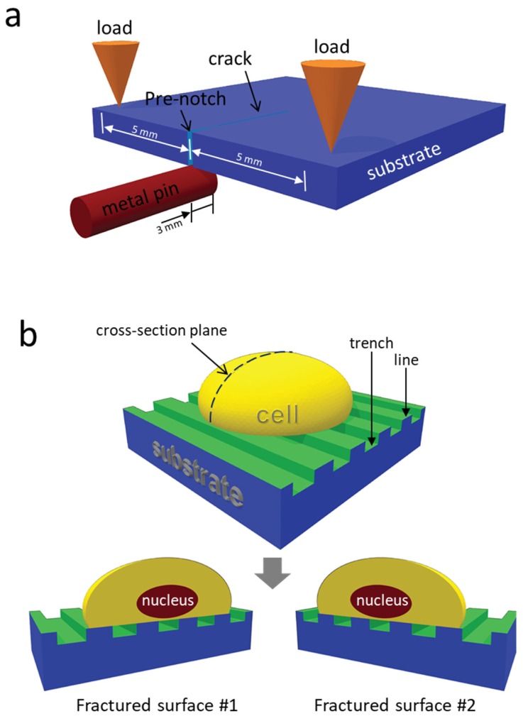
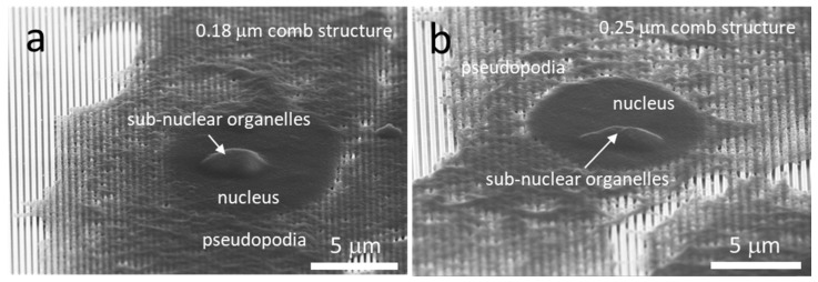
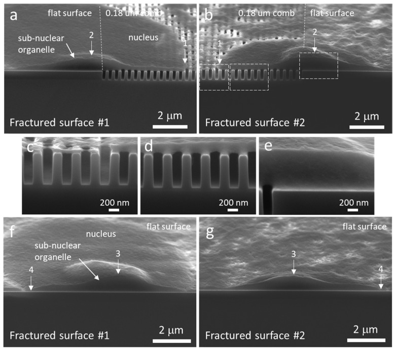
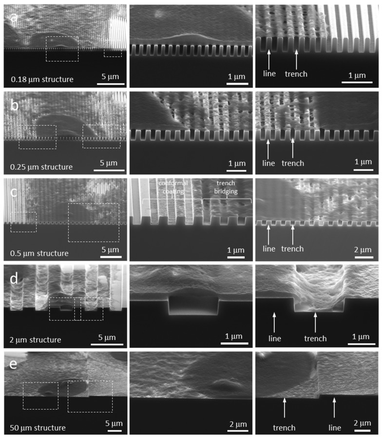
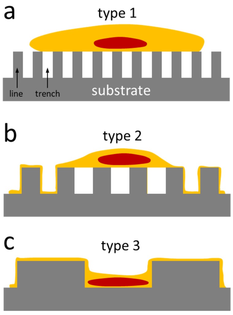
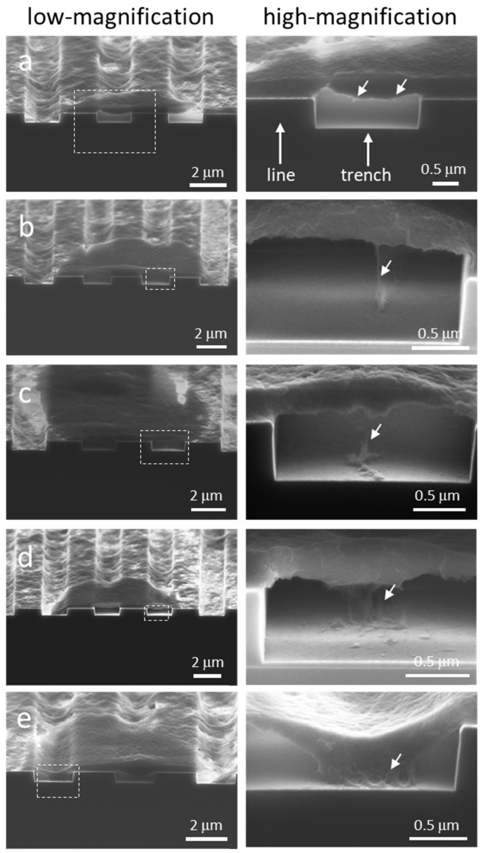
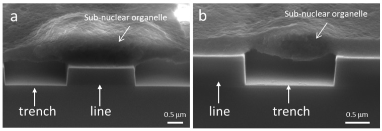
Advanced engineered surfaces can be used to direct cell behavior. These behaviors are typically characterized using either optical, atomic force, confocal, or electron microscopy; however, most microscopic techniques are generally restricted to observing what's happening on the "top" side or even the interior of the cell. Our group has focused on engineered surfaces typically reserved for microelectronics as potential surfaces to control cell behavior. These devices allow the exploration of novel substrates including titanium, tungsten, and tantalum intermixed with silicon oxide. Furthermore, these devices allow the exploration of the intricate patterning of surface materials and surface geometries i.e., trenches. Here we present two important advancements in our research: (1) the ability to split a fixed cell through the nucleus using an inexpensive three-point bend micro-cleaving technique and image 3D nanometer scale cellular components using high-resolution scanning electron microscopy; and (2) the observation of nanometer projections from the underbelly of a cell as it sits on top of patterned trenches on our devices. This application of a 3-point cleaving technique to visualize the underbelly of the cell is allowing a new understanding of how cells descend into surface cavities and is providing a new insight on cell migration mechanisms.
Keywords: adhesion; cross-sectioning; mammalian cells; morphology; nanoscale; tantalum.
Conflict of interest statement
The authors declare no conflict of interest.
Figures
Similar articles
-
Critical Design Parameters of Tantalum-Based Comb Structures to Manipulate Mammalian Cell Morphology.Materials (Basel). 2025 May 3;18(9):2099. doi: 10.3390/ma18092099. Materials (Basel). 2025. PMID: 40363602 Free PMC article.
-
Nanoscale-Textured Tantalum Surfaces for Mammalian Cell Alignment.Micromachines (Basel). 2018 Sep 13;9(9):464. doi: 10.3390/mi9090464. Micromachines (Basel). 2018. PMID: 30424397 Free PMC article.
-
Pattern-Dependent Mammalian Cell (Vero) Morphology on Tantalum/Silicon Oxide 3D Nanocomposites.Materials (Basel). 2018 Jul 28;11(8):1306. doi: 10.3390/ma11081306. Materials (Basel). 2018. PMID: 30060574 Free PMC article.
-
Single-molecule imaging of cell surfaces using near-field nanoscopy.Acc Chem Res. 2012 Mar 20;45(3):327-36. doi: 10.1021/ar2001167. Epub 2011 Oct 12. Acc Chem Res. 2012. PMID: 21992025 Review.
-
Nano-structured and functionalized surfaces for cytocompatibility improvement and bactericidal action.Biotechnol Adv. 2015 Nov 1;33(6 Pt 2):1120-9. doi: 10.1016/j.biotechadv.2015.01.001. Epub 2015 Jan 14. Biotechnol Adv. 2015. PMID: 25596482 Review.
Cited by
-
Critical Design Parameters of Tantalum-Based Comb Structures to Manipulate Mammalian Cell Morphology.Materials (Basel). 2025 May 3;18(9):2099. doi: 10.3390/ma18092099. Materials (Basel). 2025. PMID: 40363602 Free PMC article.
-
Influence of Indium (III) Chloride on Human Dermal Fibroblast Cell Adhesion on Tantalum/Silicon Oxide Nano-Composites.Materials (Basel). 2022 May 17;15(10):3577. doi: 10.3390/ma15103577. Materials (Basel). 2022. PMID: 35629602 Free PMC article.
References
-
- Coussen F., Choquet D., Sheetz M.P., Erickson H.P. Trimers of the fibronectin cell adhesion domain localize to actin filament bundles and undergo rearward translocation. J. Cell Sci. 2002;115:2581–2590. - PubMed
Grants and funding
LinkOut - more resources
Full Text Sources
