

Review
doi: 10.1038/s41556-018-0237-6.
Epub 2019 Jan 2.
Stem cell dynamics, migration and plasticity during wound healing
Affiliations
- PMID: 30602767
- PMCID: PMC7615151
- DOI: 10.1038/s41556-018-0237-6
Item in Clipboard
Review
Stem cell dynamics, migration and plasticity during wound healing
Nat Cell Biol.
2019 Jan.
Abstract
Tissue repair is critical for animal survival. The skin epidermis is particularly exposed to injuries, which necessitates rapid repair. The coordinated action of distinct epidermal stem cells recruited from various skin regions together with other cell types, including fibroblasts and immune cells, is required to ensure efficient and harmonious wound healing. A complex crosstalk ensures the activation, migration and plasticity of these cells during tissue repair.
Conflict of interest statement
The authors declare no competing interests.
Figures
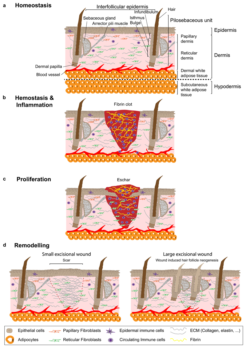
a, The skin is composed of dermis and epidermis. In the epidermis, epithelial cells are organized into a pilo-sebaceous unit, the hair follicle and its associated sebaceous glands, and the surrounding tissue, the IFE. The dermis consists of a papillary and a reticular layer located in the upper and lower part, respectively. Dermal papilla controls the hair follicle cycle and arrector pili muscle ensures its movement. The dermis includes fibroblasts, blood vessels, immune cells, sensory nerves and in its lower portion, the DWAT, which contains adipocytes. Below the skin lies the hypodermis or SWAT. b, Haemostasis and inflammation start immediately after wounding. The fibrin clot prevents further blood loss and provides a scaffold for the migration of immune, dermal and epidermal cells. c, During the proliferation phase, keratinocytes, fibroblasts and endothelial cells proliferate and migrate to the wound site and reform the ECM. d, During the remodelling phase the collagen in the dermis is remodelled and cells from earlier stages are removed. In small excisional wounds in mice, hair follicles are not regenerated and dermal scar tissue compensates for skin loss (left panel). In large excisional injuries, WIHN can be observed after complete re-epithelialization (right panel). DWAT, dermal white adipose tissue; ECM, extracellular matrix; SWAT, subcutaneous white adipose tissue; WIHN, wound induced hair follicle neogenesis.
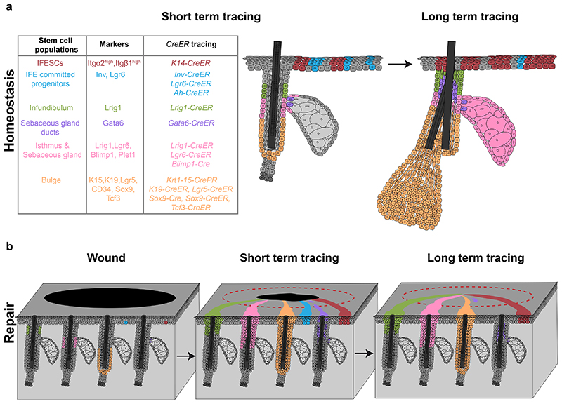
a, Skin epithelial stem cells express specific markers and can be lineage-traced with CreER mouse strains (left table). IFE stem cells and committed progenitors are located in the basal layer of the IFE and give rise to suprabasal, differentiated cells. Stem cells and committed progenitors can be traced using a K14-CreER or Inv-CreER mouse strains induced at low dose respectively. IFE committed progenitors also express Lgr6. Infundibulum stem cells are located in the upper part of the isthmus and express Lrig1. A population of sebaceous gland duct stem cells expressing Gata6 are located at the entrance of the gland but only maintains the junctional zone. Isthmus and sebaceous gland stem cells are basal cells located at the junction between the hair follicle and the gland, express Lrig1, Lgr6 and Blimp1 and give rise to the entire sebaceous gland and the isthmus. Bulge stem cells are located in the permanent lowest portion of the hair follicle, express K15, K19, Lgr5, CD34, Sox9 and Tcf3 and give rise to the entire hair follicle. b, Upon wounding, both IFESCs and committed progenitors are recruited and contribute to tissue repair. Only IFESCs will reside in the newly formed IFE long-term. Isthmus, sebaceous gland and infundibulum stem cells are recruited, contribute to IFE repair and remain long-term. Bulge stem cells are recruited to the IFE and a small proportion can remain long-term as IFESCs. Gata6+ sebaceous gland duct stem cells are recruited to the IFE, migrate suprabasally, de-differentiate and are re-established as IFESCs in the long-term. IFE, interfollicular epidermis; IFESCs, interfollicular epidermis stem cells.
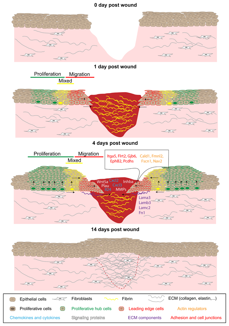
Epithelial cells start to migrate into the wound bed within 12 hours after injury. The day after wounding, IFE cells located close to the wound show an elongated shape toward the direction of the wound and are quiescent, whereas cells located at a distance start to proliferate, which leads to the establishment of a proliferative and a migrating leading edge compartment. Between these two zones, a mixed region is observed containing both migrating and proliferative cells. Four days after wounding, leading edge cells are compressed and upregulate the expression of specific genes that promote inflammation and regeneration. This gene signature is transient and disappears when the IFE is healed. IFE, interfollicular epidermis; ECM, extracellular matrix.
References
-
- Sun BK, Siprashvili Z, Khavari PA. Advances in skin grafting and treatment of cutaneous wounds. Science (New York, NY) 2014;346:941–945. - PubMed
Publication types
MeSH terms
Grants and funding
LinkOut - more resources
Full Text Sources
Other Literature Sources
Medical
