

MR imaging of intracranial solitary fibrous tumor: a retrospective study of 7 cases
- PMID: 30603014
- PMCID: PMC6306993
- DOI: 10.4314/ahs.v18i3.39
MR imaging of intracranial solitary fibrous tumor: a retrospective study of 7 cases
Abstract
Objective: To investigate the MR imaging diagnostic features of intracranial solitary fibrous tumors (ISFTs).
Materials and methods: Seven patients (mean age of 52.9 years; M:F=3:4) with histopathologically proven ISFTs were identified at our institute. Clinical presentations and pathological features were reviewed. MR Imaging findings including signal intensity, gadopentetate dimeglumine enhanced pattern, and diffusion-weighted imaging (DWI) characterization of the tumors were retrospectively evaluated.
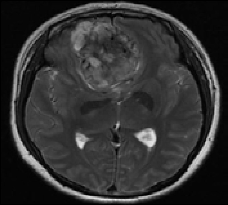
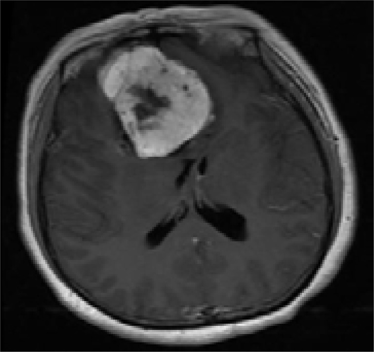
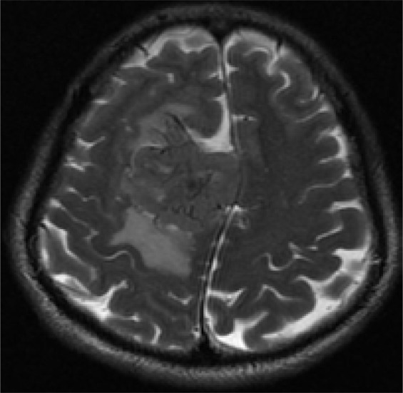
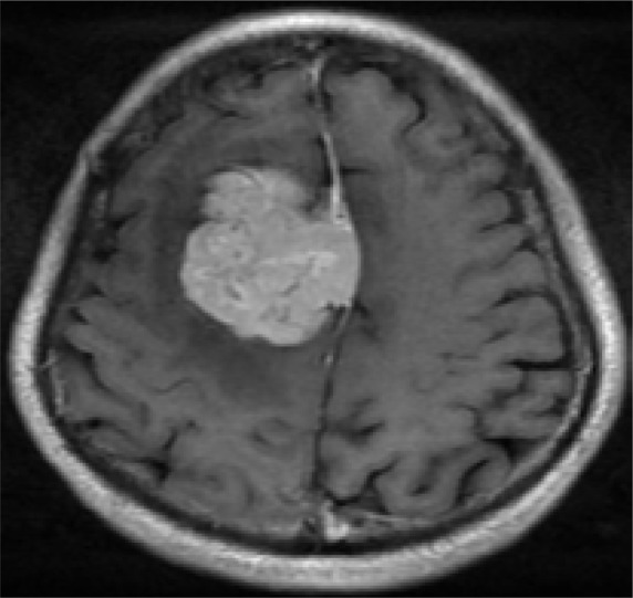
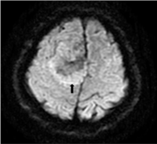
Results: Six tumors showed a multi-lobular contour. Five tumors showed heterogeneous signal intensity, and two tumors showed homogeneous signal intensity on T1WI. Low signal intensity linear, curved or interlacing lines were observed within the tumors in all seven cases. Seven tumors demonstrated moderate or strong enhancement, six showed heterogeneous enhancement, and one homogenous enhancement. All tumors showed heterogeneous signal intensity on DWI.A ring-like high signal intensity band distributed around within the tumor was noted in six cases on DWI.
Conclusion: Diagnostic evidence for ISFT on MR image includes heterogeneous signal intensity, intense enhancement of T2 signal intensity, low signal intensity lines within the tumor, heterogeneous signal intensity on DWI and a ring-like band around the tumor on DWI.
Keywords: Diffusion-weighted Imaging; Intracranial Solitary Fibrous Tumor; Magnetic resonance imaging.
Figures
Similar articles
-
Intracranial solitary fibrous tumor: imaging findings.Eur J Radiol. 2011 Nov;80(2):387-94. doi: 10.1016/j.ejrad.2010.02.016. Epub 2010 Mar 29. Eur J Radiol. 2011. PMID: 20303226
-
Evaluation of preoperative magnetic resonance imaging features and diagnostic effectiveness of grades II and III intracranial solitary fibroma.Eur J Med Res. 2024 Jul 19;29(1):377. doi: 10.1186/s40001-024-01959-5. Eur J Med Res. 2024. PMID: 39030639 Free PMC article.
-
The Value of MRI and Clinical Features in Differentiating Between Cellular and Fibrous Solitary Fibrous Tumors.AJR Am J Roentgenol. 2017 Jan;208(1):10-17. doi: 10.2214/AJR.16.16423. Epub 2016 Oct 11. AJR Am J Roentgenol. 2017. PMID: 27726413
-
Magnetic resonance imaging of thymic epithelial tumors.Crit Rev Diagn Imaging. 1996 Aug;37(3):191-259. Crit Rev Diagn Imaging. 1996. PMID: 8872410 Review.
-
MR imaging of malignant primary breast lymphoma: including diffusion-weighted imaging, histologic features, and a literature review.Jpn J Radiol. 2013 Oct;31(10):668-76. doi: 10.1007/s11604-013-0232-6. Epub 2013 Jul 12. Jpn J Radiol. 2013. PMID: 23846235 Review.
Cited by
-
Surgical Management of Craniospinal Axis Solitary Fibrous Tumors: A Single-Institution Case Series and Comprehensive Review of the Literature.Neurosurgery. 2024 Feb 1;94(2):358-368. doi: 10.1227/neu.0000000000002692. Epub 2023 Sep 25. Neurosurgery. 2024. PMID: 37747216 Free PMC article. Review.
-
Progress in infections, reproductive health and non-communicable diseases.Afr Health Sci. 2018 Sep;18(3):i-iv. doi: 10.4314/ahs.v18i3.1. Afr Health Sci. 2018. PMID: 30603019 Free PMC article. No abstract available.
-
Solitary fibrous tumor of the central nervous system invading and penetrating the skull: A case report.Oncol Lett. 2023 Jan 10;25(2):81. doi: 10.3892/ol.2023.13667. eCollection 2023 Feb. Oncol Lett. 2023. PMID: 36742362 Free PMC article.
-
A rare case of intracranial solitary fibrous tumor that is still alive after multiple surgical resections: a case report and review of the literature.Front Neurol. 2023 Jul 10;14:1201964. doi: 10.3389/fneur.2023.1201964. eCollection 2023. Front Neurol. 2023. PMID: 37492853 Free PMC article.
-
Surgical treatment for extremely rare solitary fibrous tumors of the central nervous system originating from cranial nerve VIII: new clinicopathological findings. Illustrative case.J Neurosurg Case Lessons. 2023 Aug 7;6(6):CASE23244. doi: 10.3171/CASE23244. Print 2023 Aug 7. J Neurosurg Case Lessons. 2023. PMID: 37581588 Free PMC article.
References
-
- Carneiro SS, Scheithauer BW, Nascimento AG, et al. Solitary fibrous tumor of the meninges: a lesion distinct from fibrous meningioma. A clinicopathologic and immunohistochemical study. Am J Clin Pathol. 1996;106:217–224. - PubMed
-
- Yilmaz C, Kabatas S, Ozen OI, et al. Solitary fibrous tumor. J Clin Neurosci. 2009;16:1578–1581. - PubMed
-
- Tihan T, Viglione M, Rosenblum MK, et al. Solitary fibrous tumors in the central nervous system. A clinicopathologic review of 18 cases and comparison to meningeal hemangiopericytomas. Arch Pathol Lab Med. 2003;127:432–439. - PubMed
-
- Boada M, Gomez E, Puig J, et al. Intraventricular fibrous tumor: a case report. Radiologia. 2009;51:512–515. - PubMed
MeSH terms
LinkOut - more resources
Full Text Sources
Medical