. 2019 Jan 1:5:1.
doi: 10.1038/s41421-018-0068-4. eCollection 2019.

SG formation relies on eIF4GI-G3BP interaction which is targeted by picornavirus stress antagonists

Affiliations

SG formation relies on eIF4GI-G3BP interaction which is targeted by picornavirus stress antagonists

Xiaodan Yang et al. Cell Discov. .

Abstract

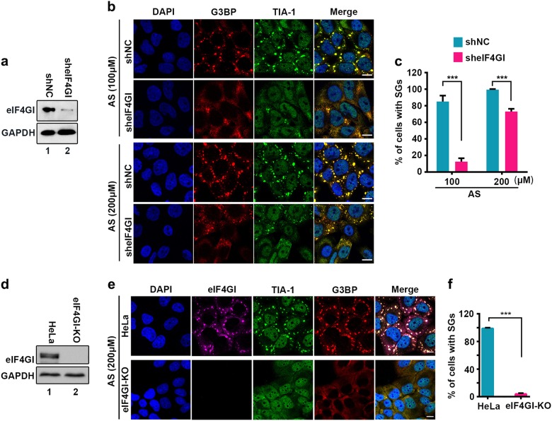
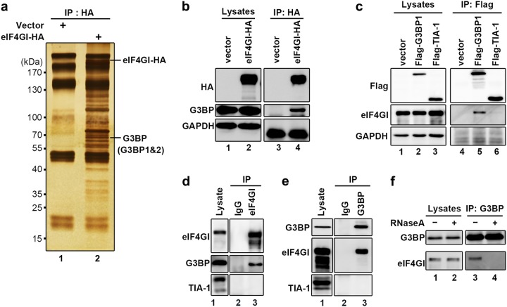
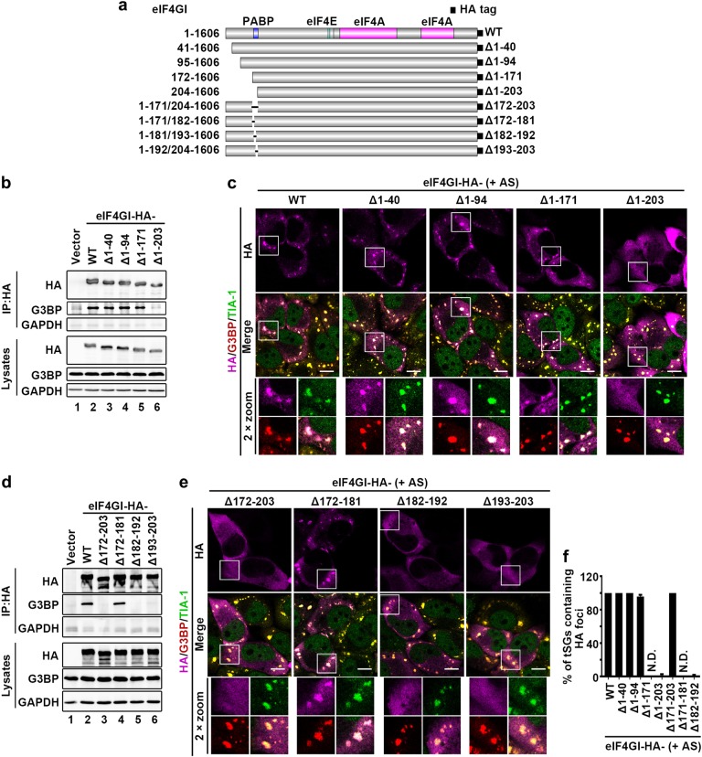
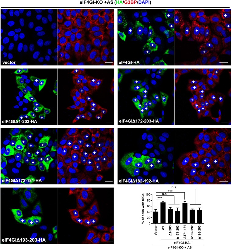
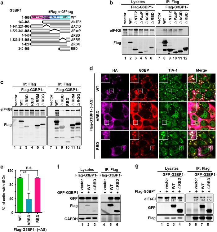
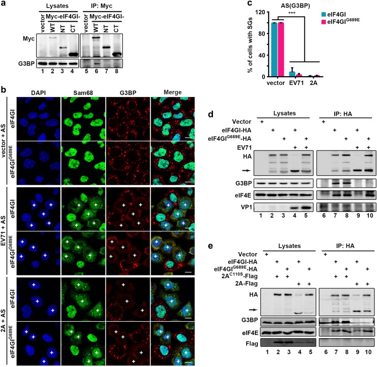
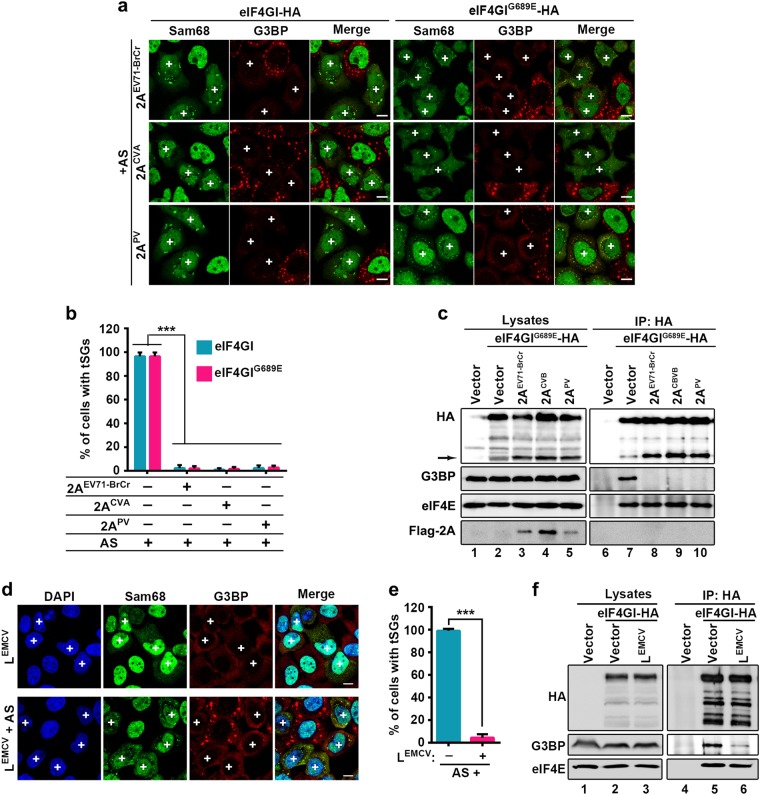
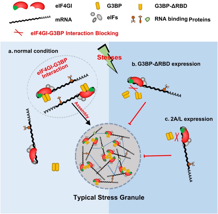
Typical stress granules (tSGs) are stalled translation pre-initiation complex aggregations in the cytoplasm, and their formation is a common consequence of translation initiation inhibition under stress. We previously found that 2A protease of picornaviruses blocks tSG formation and induces atypical SG formation, but the molecular mechanism by which 2A inhibits tSG formation remains unclear. Here, we found that eukaryotic translation initiation factor 4 gamma1 (eIF4GI) is critical for tSG formation by interacting with Ras-GTPase-activating protein SH3-domain-binding protein (G3BP), and this interaction is mediated by aa 182-203 of eIF4GI and the RNA-binding domain of G3BP. Upon eIF4GI-G3BP interaction, eIF4GI can assemble into tSGs and rescue tSG formation. Finally, we found that 2A or L protein of picornaviruses blocks tSG formation by disrupting eIF4GI-G3BP interaction. Our findings provide the first evidence that eIF4GI-G3BP interaction is indispensable for tSG formation, and 2A or L protein of picornaviruses interferes eIF4GI-G3BP interaction, thereby blocking tSG formation.


Conflict of interest statement

The authors declare that they have no conflict of interest.

Figures

Fig. 1
Fig. 1. eIF4GI is indispensable for the formation of tSGs.
ac Effects of eIF4GI KD on tSG formation. Examination of the protein level of eIF4GI in HeLa cells with KD of eIF4GI (a). Cells were treated with AS for 1 h, then fixed and stained with G3BP (red) and TIA-1 (green). IF (b) and quantitation (c) analysis of cells with tSGs (G3BP served as a marker). n = 3, 300 cells/assay were counted, mean ± SD; ***p < 0.001. df Effects of eIF4GI KO on tSG formation. Examination of the protein level of eIF4GI in HeLa cells with KO of eIF4GI (HeLa-eIF4GI-KO cells) (d). Cells were treated with AS for 1 h and then fixed and stained with eIF4GI (purple), G3BP (red), and TIA-1 (green). IF (e) and quantitation (f) analysis of cells with tSGs (G3BP served as a marker). n = 3, 300 cells/assay were counted, mean ± SD; ***p < 0.001. Scale bars, 10 μm. See also Supplementary Fig. S1
Fig. 2
Fig. 2. eIF4GI interacts with G3BP.
ab HeLa cells were transfected with eIF4GI-HA for 24 h, and cell lysates were subjected to IP/MS with anti-HA antibody. Immunoprecipitates were separated via SDS-PAGE and analyzed via silver staining (a) or WB (b).The anti-G3BP antibody, which used to detect endogenous G3BP, can recognize both G3BP1 and G3BP2. c To detect the interaction of G3BP or TIA-1 with endogenous eIF4GI, HeLa cells expressing Flag-tagged G3BP1 or TIA-1 were lysed and subjected to IP with anti-Flag antibody, followed by WB to resolve the immune complexes. de Interaction examination between endogenous eIF4GI and G3BP (TIA-1 was used as negative control). HeLa cells were lysed and subjected to IP with antibodies against eIF4GI (d) or G3BP (e). Lysates and immunoprecipitates were resolved via WB with indicated antibodies. f HeLa cells were lysed and treated with RNaseA ( + ) or mock-treated (−) before IP with anti-G3BP antibody, followed by detection of eIF4GI and G3BP via Western blots. See also Supplementary Fig. S2
Fig. 3
Fig. 3. eIF4GI-G3BP interaction is required for eIF4GI assembly into tSGs.
a Graphic description of eIF4GI and its mutants. bf eIF4GI-HA and its mutants were expressed as indicated. Cell lysates were subjected to IP and analyzed as in Fig. 2b (b and d). Cells were treated with 200 μM AS for 1 h, then fixed and stained with HA (purple), G3BP (red), and TIA-1 (green). G3BP and TIA-1 served as indicators of tSGs (c and e). Quantitation of SGs containing indicated HA foci in c and e (f). n = 3, 300 cells/assay were counted, mean ± SD, N.D., not detectable. Scale bars, 10 μm. Supplementary Fig. S3
Fig. 4
Fig. 4. tSG formation rescue assays in HeLa-eIF4GI-KO cells.
HeLa-eIF4GI-KO cells were transfected with HA-tagged eIF4GI or its mutants as indicated for 24 h, treated with AS for 1 h, and then subjected to IF assay. Cells were stained with HA (green), G3BP (red), and DAPI (blue). “ + ” indicates cells expressing HA-tagged protein. Quantitation of HA-positive cells with G3BP-marked tSGs (bottom and right panel). For “Vector” column, the overall percentage of cells with SGs was calculated. n = 3, 400 cells/condition were counted, mean ± SD; ***p < 0.001, n.s. no statistical significance. Scale bars, 10 μm
Fig. 5
Fig. 5. RBD of G3BP is required for eIF4GI-G3BP interaction.
a Graphic description of G3BP and its mutants. b and c HeLa cells were transfected as indicated, and cell lysates were processed as in Fig. 2c. de HeLa cells were transfected as indicated, treated with AS (200 μM, 1 h), and stained with TIA-1 (green) and G3BP (red) to visualize the tSGs and flag (purple) to visualize the expression of G3BP and its mutants (d). Quantitation of Flag-positive cells with TIA-1-marked tSGs (e). n = 3, 240 cells/condition were counted, mean ± SD; ***p < 0.001, n.s. no statistical significance. fg HeLa cells transfected as indicated, and cell lysates were processed as in (b). Scale bars, 10 μm
Fig. 6
Fig. 6. EV71 and 2A block tSG formation by disrupting eIF4GI-G3BP interaction.
a HeLa cells were transfected as indicated. Cell lysates were subjected to IP with an anti-Myc antibody. b eIF4GI-HA- or eIF4GIG689E-HA-HeLa cells were transfected with vector or 2A for 24h, or infected with EV71 (MOI = 10) for 5 h, then treated with 200 μM AS for 1 h, and stained with Sam68 (green) and G3BP (red). Sam68 served as an indicator of EV71 infection or 2A expression. “ + ” indicates EV71-infected or 2A-expressing cells. c Quantitation analysis of vector-transfected, EV71-infected, or 2A-expressing cells with tSGs in (b). n = 3, 240 cells/condition were counted, mean ± SD; ***p < 0.001. d eIF4GI-HA- or eIF4GIG689E-HA-HeLa cells were infected with EV71 as indicated, and cell lysates were subjected to IP with an anti-HA antibody. The bound proteins were analyzed via WB. VP1 indicated EV71 infection. Arrow indicates eIF4GI cleavage products. e eIF4GI-HA- or eIF4GIG689E-HA-HeLa cells were transfected with Flag-tagged 2A or 2AC110S for 24 h and analyzed as in (d). Scale bars, 10 μm
Fig. 7
Fig. 7. Disruption of eIF4GI-G3BP interaction by 2A/L to block tSG formation is common among picornaviruses.
a and b eIF4GI-HA- and eIF4GIG689E-HA-HeLa cells were transfected with 2A of EV71-BrCr, CVA, or PV for 24 h, and cells were processed as in Fig. 6a (a) and quantified as in Fig. 6b (b). (c) eIF4GIG689E-HA-HeLa cells were transfected with 2A of EV71-BrCr, CVA, or PV for 24 h and analyzed as in Fig. 6d. (d) HeLa cells were transfected with LEMCV for 24 h, then treated with 200 μM AS or not. Cells were stained with Sam68 (green) to visualize expression of LEMCV and G3BP (red) to visualize tSGs. e Quantitation of LEMCV-expressing cells with tSGs in (d). n = 3, 240 cells/condition were counted, mean ± SD; ***p < 0.001. f eIF4GI-HA-HeLa cells were transfected with LEMCV for 24 h, and cell lysates were subjected to IP as in (b). “ + ” indicates 2A or LEMCV-expressing cells. Scale bars, 10 μm
Fig. 8
Fig. 8. Models of the regulation of tSG assembly by eIF4GI-G3BP interaction.
a In normal condition, stresses promote the interaction between eIF4GI and G3BP, which leads to the assembly of eIF4GI into tSGs and the rescue of tSG formation. b-c The expression of G3BP-ΔRBD (b) or 2A/L (c) interferes eIF4GI-G3BP interaction to block tSG assembly

Similar articles

Cited by

References

    1. Anderson P, Kedersha N. Stress granules: the Tao of RNA triage. Trends Biochem. Sci. 2008;33:141–150. doi: 10.1016/j.tibs.2007.12.003. - DOI - PubMed
    1. Kedersha N, et al. Dynamic shuttling of TIA-1 accompanies the recruitment of mRNA to mammalian stress granules. J. Cell Biol. 2000;151:1257–1268. doi: 10.1083/jcb.151.6.1257. - DOI - PMC - PubMed
    1. Tourriere H, et al. The RasGAP-associated endoribonuclease G3BP assembles stress granules. J. Cell Biol. 2003;160:823–831. doi: 10.1083/jcb.200212128. - DOI - PMC - PubMed
    1. Gilks N, et al. Stress granule assembly is mediated by prion-like aggregation of TIA-1. Mol. Biol. Cell. 2004;15:5383–5398. doi: 10.1091/mbc.e04-08-0715. - DOI - PMC - PubMed
    1. Burgess HM, et al. Nuclear relocalisation of cytoplasmic poly(A)-binding proteins PABP1 and PABP4 in response to UV irradiation reveals mRNA-dependent export of metazoan PABPs. J. Cell Sci. 2011;124:3344–3355. doi: 10.1242/jcs.087692. - DOI - PMC - PubMed