. 2016 Nov 1;7(4):342-351.
doi: 10.1007/s13340-016-0290-y. eCollection 2016 Dec.

M2 macrophages in metabolism

Affiliations

M2 macrophages in metabolism

Shiho Fujisaka et al. Diabetol Int. .

Abstract

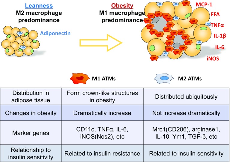
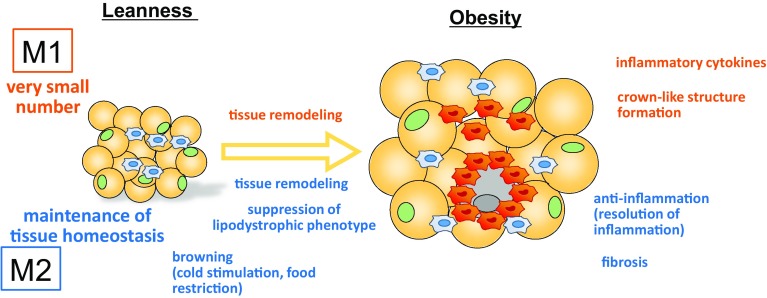
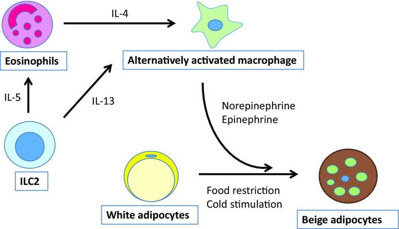
Adipose tissue not only functions as the major energy-storing tissue, but also functions as an endocrine organ that regulates systemic metabolism by releasing various hormones called adipokines. Macrophages play a critical role in maintaining adipocyte health in a lean state and in remodeling during the progression of obesity. Large numbers of classically activated (M1) macrophages accumulate in adipose tissue as adipocytes become larger because of excessive energy conditions, and they adversely affect insulin resistance by triggering local and systemic inflammation. In contrast, alternatively activated (M2) macrophages seem to maintain the health of adipose tissues in a lean state. In addition, they play a role in adapting to excess energy states, because M2 macrophage dysfunction caused by genetic disruption of the M2 gene results in metabolic disorders under high-fat-fed conditions that are probably attributable to their anti-inflammatory functions. Nonetheless, how M2 macrophages contribute to maintaining the health of adipose tissue and therefore to insulin sensitivity is largely unknown. In this article, we review the literature on the role of M1 and M2 macrophages in metabolism, with a special focus on the role of M2 macrophages in adipose tissue. Likewise, we raise topics of M2 macrophages in non-adipose tissues to expand our understanding of macrophage heterogeneity.

Keywords: Adipose tissue; Beiging; Insulin resistance; Macrophage; Obesity.


Conflict of interest statement

The authors declare the following financial interests: AstraZeneca K.K., Merck & Co., Inc., Medical Review Co., Ltd., Takeda Pharmaceutical Co., Ltd., Mitsubishi Tanabe Pharma, Novo Nordisk Pharma, Ltd., Kowa Pharmaceutical Co., Ltd., Astellas Pharma Inc., and Fuji Chemical Industries Co., Ltd.This article does not contain any studies with human or animal subjects performed by any of the authors.

Figures

Fig. 1
Fig. 1
The heterogeneity of macrophages in the development of adipose tissue inflammation. In the lean state, M2 ATMs predominate. As obesity progresses, monocytes accumulate in adipose tissue and differentiate into inflammatory M1 or anti-inflammatory M2 macrophages. Since the number of M1 ATMs increases dramatically, whereas there is only a modest increase in the number of M2 ATMs, M1 ATMs predominate in obese adipose tissue. M1 ATMs produce inflammatory cytokines and promote insulin resistance. M2 ATMs, on the other hand, secrete anti-inflammatory cytokines, including IL-10 and TGF-β, and suppress inflammation, suggesting that M2 ATMs are associated with insulin sensitivity
Fig. 2
Fig. 2
Functional differences between M1 ATMs and M2 ATMs. In the lean state, the major ATMs are the M2 phenotype, and they play roles in maintaining tissue homeostasis. During cold stimulation and caloric restriction, M2 ATMs are involved in the browning of subQ. As obesity progresses both M1 and M2 ATMs become involved in tissue remodeling. M1 ATMs accumulate around necrotic adipocytes and form crown-like structures (CLSs), which further contribute to polarization into the M1 phenotype by activating HIF-1α. By contrast, M2 ATMs somehow prevent the development of the lipodystrophic phenotype, and they may play a role in suppressing inflammatory responses induced by M1 ATMs. In some situations, M2 ATMs induce fibrosis, thereby aggravating insulin resistance
Fig. 3
Fig. 3
A simplified view of the relationship between M2 ATMs and other cells in beiging. ILC2s secrete IL-5, which activates eosinophils, and they produce IL-4 in response. IL-4 induces M2 polarization. M2 macrophages produce norepinephrine, which in turn promotes differentiation toward beige adipocytes

References

    1. Hoeffel G, Wang Y, Greter M, See P, Teo P, Malleret B, Leboeuf M, Low D, Oller G, Almeida F, et al. Adult Langerhans cells derive predominantly from embryonic fetal liver monocytes with a minor contribution of yolk sac-derived macrophages. J Exp Med. 2012;209(6):1167–1181. doi: 10.1084/jem.20120340. - DOI - PMC - PubMed
    1. Wynn TA, Chawla A, Pollard JW. Macrophage biology in development, homeostasis and disease. Nature. 2013;496(7446):445–455. doi: 10.1038/nature12034. - DOI - PMC - PubMed
    1. Schulz C, Gomez Perdiguero E, Chorro L, Szabo-Rogers H, Cagnard N, Kierdorf K, Prinz M, Wu B, Jacobsen SE, Pollard JW, et al. A lineage of myeloid cells independent of Myb and hematopoietic stem cells. Science. 2012;336(6077):86–90. doi: 10.1126/science.1219179. - DOI - PubMed
    1. Mantovani A, Sica A, Sozzani S, Allavena P, Vecchi A, Locati M. The chemokine system in diverse forms of macrophage activation and polarization. Trends Immunol. 2004;25(12):677–686. doi: 10.1016/j.it.2004.09.015. - DOI - PubMed
    1. Takeuchi O, Akira S. Signaling pathways activated by microorganisms. Curr Opin Cell Biol. 2007;19(2):185–191. doi: 10.1016/j.ceb.2007.02.006. - DOI - PubMed

LinkOut - more resources