

Diffusion-weighted magnetic resonance imaging in the pancreas of fulminant type 1 diabetes
- PMID: 30603375
- PMCID: PMC6224878
- DOI: 10.1007/s13340-018-0355-1
Diffusion-weighted magnetic resonance imaging in the pancreas of fulminant type 1 diabetes
Abstract
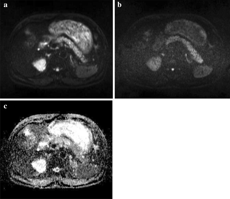
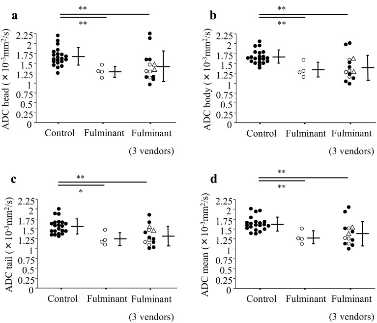
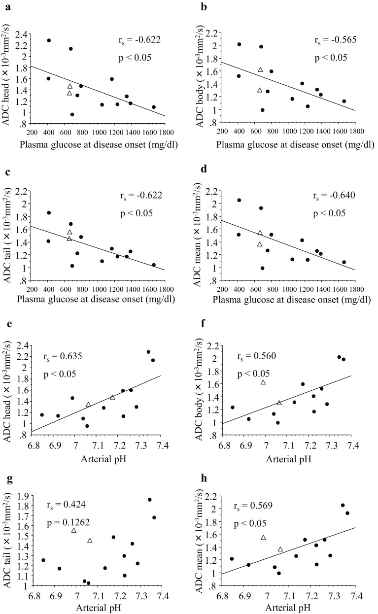
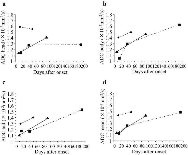
Abrupt disease onset and severe metabolic disorders are main characteristics of fulminant type 1 diabetes. Diffusion-weighted magnetic resonance imaging (DWI) is an imaging technique that reflects restricted diffusion in organs and can detect mononuclear cell infiltration into the pancreas at the onset of the disease. Fourteen patients with fulminant type 1 diabetes who underwent abdominal magnetic resonance imaging were recruited for the measurement of apparent diffusion coefficient (ADC) values of the pancreas that were compared with those of 21 non-diabetic controls. The ADC values of all parts of the pancreas were significantly lower in fulminant type 1 diabetes than in controls (head, 1.424 ± 0.382 × 10⁻³ vs. 1.675 ± 0.227 × 10⁻³ mm2/s; body, 1.399 ± 0.317 × 10⁻³ vs. 1.667 ± 0.170 × 10⁻³ mm2/s; tail, 1.336 ± 0.247 × 10⁻³ vs. 1.561 ± 0.191 × 10⁻³ mm2/s; mean, 1.386 ± 0.309 × 10⁻³ vs. 1.634 ± 0.175 × 10⁻³ mm2/s) (p < 0.01). The best cut-off value indicated that the sensitivity was 86% and the specificity was 71% when using DWI, which was also efficient in two atypical patients with fulminant type 1 diabetes without elevated levels of exocrine pancreatic enzymes or with high HbA1c levels due to the preexistence of type 2 diabetes. The ADC values were significantly correlated to plasma glucose levels and arterial pH, and tended to increase with the lapse of time. DWI may be an additional tool for making an efficient diagnosis of fulminant type 1 diabetes.
Keywords: Apparent diffusion coefficient; Diffusion-weighted imaging; Fulminant; Magnetic resonance imaging; Type 1 diabetes.
Conflict of interest statement
The authors declare no conflicts of interest.All procedures followed were in accordance with the ethical standards of the responsible committee on human experimentation (institutional and national) and with the Helsinki Declaration of 1964 and later versions. Opt-out opportunities are provided to study subjects.
Figures
Similar articles
-
Fulminant type 1 diabetes: recent research progress and future prospects.Diabetol Int. 2020 Sep 16;11(4):336-341. doi: 10.1007/s13340-020-00466-2. eCollection 2020 Oct. Diabetol Int. 2020. PMID: 33088640 Free PMC article. Review.
-
Non-breath-hold high b-value diffusion-weighted MRI with parallel imaging technique: apparent diffusion coefficient determination in normal abdominal organs.Diagn Interv Radiol. 2008 Jun;14(2):83-7. Diagn Interv Radiol. 2008. PMID: 18553281
-
Apparent diffusion coefficient measurements of the pancreas, pancreas carcinoma, and mass-forming focal pancreatitis.Acta Radiol. 2012 Mar 1;53(2):135-9. doi: 10.1258/ar.2011.100252. Epub 2012 Jan 19. Acta Radiol. 2012. PMID: 22262868
-
Diffusion-weighted magnetic resonance imaging without bowel preparation for detection of ulcerative colitis.World J Gastroenterol. 2015 Sep 7;21(33):9785-92. doi: 10.3748/wjg.v21.i33.9785. World J Gastroenterol. 2015. PMID: 26361426 Free PMC article.
-
Diffusion-weighted magnetic resonance imaging of the pancreas.Top Magn Reson Imaging. 2009 Feb;20(1):43-7. doi: 10.1097/RMR.0b013e3181b48667. Top Magn Reson Imaging. 2009. PMID: 19687725 Review.
Cited by
-
Cross-sectional imaging of the pancreas in diabetes.Abdom Radiol (NY). 2024 Jun;49(6):2116-2124. doi: 10.1007/s00261-024-04310-y. Epub 2024 Apr 1. Abdom Radiol (NY). 2024. PMID: 38557767 Free PMC article. Review.
-
New-onset atypical fulminant type 1 diabetes after COVID-19 vaccination: A case report.Clin Case Rep. 2022 Oct 17;10(10):e6473. doi: 10.1002/ccr3.6473. eCollection 2022 Oct. Clin Case Rep. 2022. PMID: 36267825 Free PMC article.
-
Immune checkpoint inhibitor-related diabetes mellitus associated with high signal intensity in diffusion-weighted magnetic resonance imaging of the pancreas at an early clinical stage.Hormones (Athens). 2025 Jun;24(2):569-574. doi: 10.1007/s42000-025-00629-3. Epub 2025 Jan 17. Hormones (Athens). 2025. PMID: 39821892 Free PMC article.
-
Fulminant type 1 diabetes: recent research progress and future prospects.Diabetol Int. 2020 Sep 16;11(4):336-341. doi: 10.1007/s13340-020-00466-2. eCollection 2020 Oct. Diabetol Int. 2020. PMID: 33088640 Free PMC article. Review.
References
-
- Expert Committee on the Diagnosis and Classification of Diabetes Mellitus Report of the expert committee on the diagnosis and classification of diabetes mellitus. Diabetes Care. 2003;26(Suppl 1):S5–S20. - PubMed
-
- Alberti KG, Zimmet PZ. Definition, diagnosis and classification of diabetes mellitus and its complications. Part 1: diagnosis and classification of diabetes mellitus provisional report of a WHO consultation. Diabet Med. 1998;15:539–553. doi: 10.1002/(SICI)1096-9136(199807)15:7<539::AID-DIA668>3.0.CO;2-S. - DOI - PubMed
LinkOut - more resources
Full Text Sources