Review
. 2017 May 18;14(3):201-210.
doi: 10.1007/s13770-017-0026-5. eCollection 2017 Jun.

Mesenchymal Stem Cell-Based Therapies against Podocyte Damage in Diabetic Nephropathy

Affiliations
Review

Mesenchymal Stem Cell-Based Therapies against Podocyte Damage in Diabetic Nephropathy

Manizheh Khalilpourfarshbafi et al. Tissue Eng Regen Med. .

Abstract

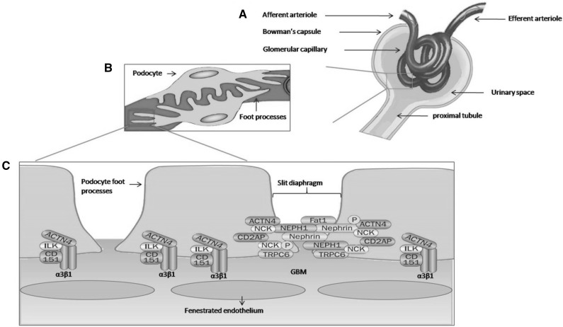
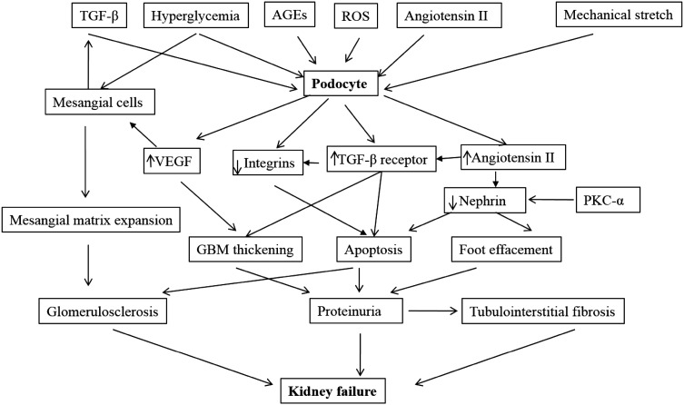
Injury to podocytes is an early event in diabetic nephropathy leading to proteinuria with possible progression to end-stage renal failure. The podocytes are unique and highly specialized cells that cover the outer layer of kidney ultra-filtration barrier and play an important role in glomerular function. In the past few decades, adult stem cells, such as mesenchymal stem cells (MSCs) with a regenerative and differentiative capacity have been extensively used in cell-based therapies. In addition to their capability for regeneration and differentiation, MSCs contributes to their milieu by paracrine action of a series of growth factors via antiapoptotic, mitogenic and other cytokine actions that actively participate in treatment of podocyte damage through prevention of podocyte effacement, detachment and apoptosis. It is hoped that novel stem cell-based therapies will be developed in the future to prevent podocyte injury, thereby reducing the burden of kidney disease.

Keywords: Diabetic nephropathy; Glomerular function; Kidney ultrafiltration; Mesenchymal stem cells; Podocyte damage.


Conflict of interest statement

The authors have declared that no conflict of interest.There are no animal experiments carried out for this article.

Figures

Fig. 1
Fig. 1
Structure of the glomerular filtration barrier. A) Fluids from blood in the glomerulus are collected in the Bowman’s capsule that empties into proximal tubules. B) The glomerular filtration barrier consist of three layers: the innermost fenestrated endothelium, the glomerular basement membrane (GBM), and the podocyte layer. C) The foot process of neighboring podocytes interconnected by several slit diaphragm molecules. Proteins that anchor foot processes to the GBM (α3β1 integrin, α-actinin-4 [ACTN4], integrin linked kinase [ILK], and the tetraspanin CD151) and those associated with the slit diaphragm (nephrin, NEPH1, podocin [P], Fat1, ACTN4, the adaptor protein NCK, CD2-associated protein [CD2AP], and transient receptor potential cation channel 6 [TRPC6]) are important in maintaining of filtration barrier
Fig. 2
Fig. 2
Podocyte damage in diabetic nephropathy. In diabetic condition, metabolic factors (TGF-β, glycated proteins, hyperglycemia, ROS, Angiotensin II (Ang II) and hemodynamic factors (via mechanical stretch) lead to increased VEGF and Ang II production by podocytes. Podocyte-derived VEGF leads to overproduction of matrix in mesangial cells and diabetic glomerulosclerosis. Chronic hyperglycemia and advanced glycation end products lead to TGF-β secretion in mesangial cells and expression of TGF-β R’ in podocytes. The latent TGF-β complex may be stored in mesangial matrix and then localized to the podocyte surface via activation by Ang II. The TGF-β type II receptor interaction stimulates the over production of extracellular matrix by podocyte and mesangium (leading to GBM thickening and mesangial matrix expansion). Over expression of TGF-β and mechanical stretch suppress α3β1 integrin and leads to decreased podocyte adhesion and apoptosis. In response to high glucose concentration, protein kinase C-alpha (PKC-α) expression increase which promotes nephrin endocytosis. This leads to a decline in nephrin surface expression and eventually contributes to proteinuria. Worsening proteinuria coupled glomerulosclerosis and tubulointerstitial fibrosis, leads to progressive renal insufficiency

References

    1. Sarje SK, Ghiware NB, Kawade RM, Gunjkar VN, Vadvalkar SM. Association of chronic complications of type 2 diabetes with the biochemical and physical estimations in subjects attending single visit screening for complications. Int J Res Pharm Chem. 2013;3:842–845.
    1. Hajiaghaalipour F, Khalilpourfarshbafi M, Arya A. Modulation of glucose transporter protein by dietary flavonoids in type 2 diabetes mellitus. Int J Biol Sci. 2015;11:508–524. doi: 10.7150/ijbs.11241. - DOI - PMC - PubMed
    1. Ezquer FE, Ezquer ME, Parrau DB, Carpio D, Yanez AJ, Conget PA. Systemic administration of multipotent mesenchymal stromal cells reverts hyperglycemia and prevents nephropathy in type 1 diabetic mice. Biol Blood Marrow Transpl J Am Soc Blood Marrow Transpl. 2008;14:631–640. doi: 10.1016/j.bbmt.2008.01.006. - DOI - PubMed
    1. Bakris GL, Williams M, Dworkin L, Elliott WJ, Epstein M, Toto R, et al. Preserving renal function in adults with hypertension and diabetes: a consensus approach. National Kidney Foundation Hypertension and Diabetes Executive Committees Working Group. Am J kidney Dis Off J Natl Kidney Found. 2000;36:646–661. doi: 10.1053/ajkd.2000.16225. - DOI - PubMed
    1. Wolf G, Chen S, Ziyadeh FN. From the periphery of the glomerular capillary wall toward the center of disease: podocyte injury comes of age in diabetic nephropathy. Diabetes. 2005;54:1626–1634. doi: 10.2337/diabetes.54.6.1626. - DOI - PubMed

LinkOut - more resources