

Heparin/Collagen 3D Scaffold Accelerates Hepatocyte Differentiation of Wharton's Jelly-Derived Mesenchymal Stem Cells
- PMID: 30603500
- PMCID: PMC6171614
- DOI: 10.1007/s13770-017-0048-z
Heparin/Collagen 3D Scaffold Accelerates Hepatocyte Differentiation of Wharton's Jelly-Derived Mesenchymal Stem Cells
Abstract
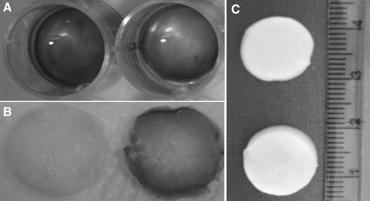
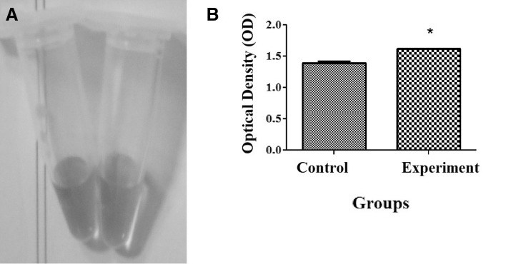
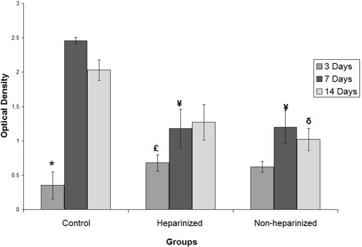
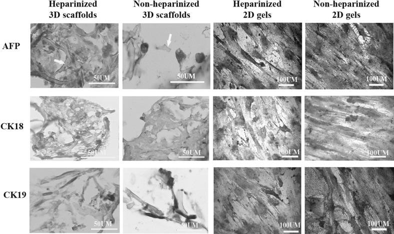
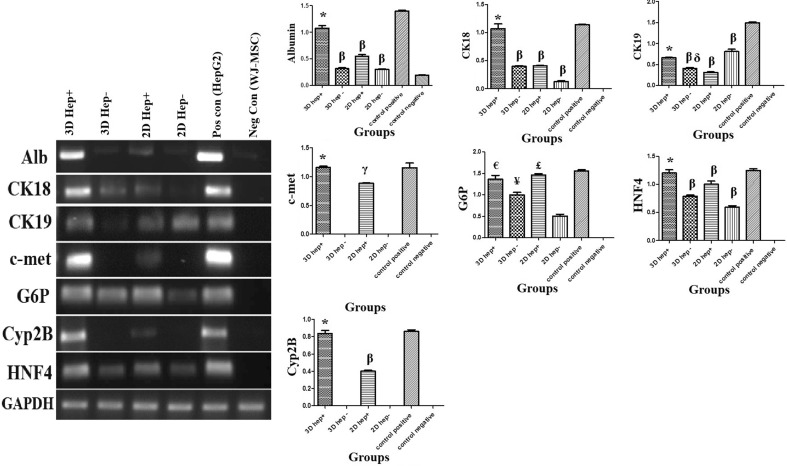
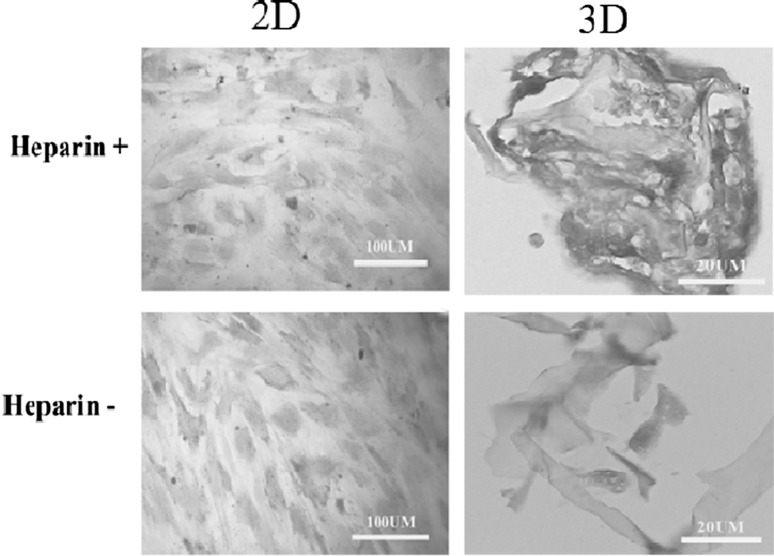
Both mature and stem cell-derived hepatocytes lost their phenotype and functionality under conventional culture conditions. However, the 3D scaffolds containing the main extracellular matrix constitutions, such as heparin, may provide appropriate microenvironment for hepatocytes to be functional. The current study aimed to investigate the efficacy of the differentiation capability of hepatocytes derived from human Wharton's jelly mesenchymal stem cells (WJ-MSCs) in 3D heparinized scaffold. In this case, the human WJ-MSCs were cultured on the heparinized and non-heparinized 2D collagen gels or within 3D scaffolds in the presence of hepatogenic medium. Immunostaining was performed for anti-alpha fetoprotein, cytokeratin-18 and -19 antibodies. RT-PCR was performed for detection of hepatic nuclear factor-4 (HNF-4), albumin, cytokeratin-18 and -19, glucose-6-phosphatase (G6P), c-met and Cyp2B. The results indicated that hepatogenic media induced the cells to express early liver-specific markers including HNF4, albumin, cytokeratin-18 and 19 in all conditions. The cells cultured on both heparinized culture conditions expressed late liver-specific markers such as G6P and Cyp2B as well. Besides, the hepatocytes differentiated in 3D heparinized scaffolds stored more glycogen that indicated they were more functional. Non-heparinized 2D gel was the superior condition for cholangiocyte differentiation as indicated by higher levels of cytokeratin 19 expression. In conclusion, the heparinized 3D scaffolds provided a microenvironment to mimic Disse space. Therefore, 3D heparinized collagen scaffold can be suggested as a good vehicle for hepatocyte differentiation.
Keywords: Collagen type I; Gel; Heparin; Hepatocytes; Scaffold.
Conflict of interest statement
There is no conflict of interest.The experimental design was approved by ethic committee of Shiraz University of Medical Sciences (IR.SUMS.REC.1395.S803).
Figures
Similar articles
-
Comparison of the Expression of Hepatic Genes by Human Wharton's Jelly Mesenchymal Stem Cells Cultured in 2D and 3D Collagen Culture Systems.Iran J Med Sci. 2016 Jan;41(1):28-36. Iran J Med Sci. 2016. PMID: 26722142 Free PMC article.
-
Comparison of hepatic nuclear factor-4 expression in two- and three-dimensional culture of Wharton's jelly-derived cells exposed to hepatogenic medium.Rom J Morphol Embryol. 2015;56(4):1365-70. Rom J Morphol Embryol. 2015. PMID: 26743282
-
The influence of fibroblast growth factor 4 on hepatogenic capacity of Wharton's jelly mesenchymal stromal cells.Rom J Morphol Embryol. 2015;56(3):1043-50. Rom J Morphol Embryol. 2015. PMID: 26662137
-
Wharton's Jelly Mesenchymal Stem Cell: Various Protocols for Isolation and Differentiation of Hepatocyte-Like Cells; Narrative Review.Iran J Med Sci. 2019 Nov;44(6):437-448. doi: 10.30476/ijms.2019.44952. Iran J Med Sci. 2019. PMID: 31875078 Free PMC article. Review.
-
Application potential of mesenchymal stem cells derived from Wharton's jelly in liver tissue engineering.Biomed Mater Eng. 2015;25(1 Suppl):137-43. doi: 10.3233/BME-141232. Biomed Mater Eng. 2015. PMID: 25538064 Review.
Cited by
-
Human umbilical cord mesenchymal stem cells ameliorate liver fibrosis in vitro and in vivo: From biological characteristics to therapeutic mechanisms.World J Stem Cells. 2019 Aug 26;11(8):548-564. doi: 10.4252/wjsc.v11.i8.548. World J Stem Cells. 2019. PMID: 31523373 Free PMC article. Review.
-
Transplantation of mesenchymal stem cells and their derivatives effectively promotes liver regeneration to attenuate acetaminophen-induced liver injury.Stem Cell Res Ther. 2020 Feb 27;11(1):88. doi: 10.1186/s13287-020-01596-9. Stem Cell Res Ther. 2020. PMID: 32106875 Free PMC article. Review.
-
Mesenchymal Stem Cells as a Promising Cell Source for Integration in Novel In Vitro Models.Biomolecules. 2020 Sep 10;10(9):1306. doi: 10.3390/biom10091306. Biomolecules. 2020. PMID: 32927777 Free PMC article. Review.
-
Ameliorating effect of encapsulated hepatocyte-like cells derived from umbilical cord in high mannuronic alginate scaffolds on acute liver failure in rats.Iran J Basic Med Sci. 2018 Sep;21(9):928-935. doi: 10.22038/IJBMS.2018.27928.6847. Iran J Basic Med Sci. 2018. PMID: 30524693 Free PMC article.
-
Heparin and Derivatives for Advanced Cell Therapies.Int J Mol Sci. 2021 Nov 7;22(21):12041. doi: 10.3390/ijms222112041. Int J Mol Sci. 2021. PMID: 34769471 Free PMC article. Review.
References
LinkOut - more resources
Full Text Sources
Other Literature Sources
Miscellaneous