Review
. 2017 Nov 20;14(6):653-665.
doi: 10.1007/s13770-017-0093-7. eCollection 2017 Dec.

Current Understanding of Stem Cell and Secretome Therapies in Liver Diseases

Affiliations
Review

Current Understanding of Stem Cell and Secretome Therapies in Liver Diseases

Dongkyu Kim et al. Tissue Eng Regen Med. .

Abstract

Liver failure is one of the main risks of death worldwide, and it originates from repetitive injuries and inflammations of liver tissues, which finally leads to the liver cirrhosis or cancer. Currently, liver transplantation is the only effective treatment for the liver diseases although it has a limitation due to donor scarcity. Alternatively, cell therapy to regenerate and reconstruct the damaged liver has been suggested to overcome the current limitation of liver disease cures. Several transplantable cell types could be utilized for recovering liver functions in injured liver, including bone marrow cells, mesenchymal stem cells, hematopoietic stem cells, macrophages, and stem cell-derived hepatocytes. Furthermore, paracrine effects of transplanted cells have been suggested as a new paradigm for liver disease cures, and this application would be a new strategy to cure liver failures. Therefore, here we reviewed the current status and challenges of therapy using stem cells for liver disease treatments.

Keywords: Liver failure; Liver regeneration; Secretome; Stem cell transplantation.


Conflict of interest statement

The authors declare that they have no competing interests.This review article does not contain any studies with human or animal subjects performed by any of the authors.

Figures
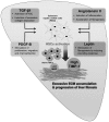
Fig. 1
Fig. 1
Mechanism for development and progression of liver fibrosis. Continuous damages on liver tissue result in the secretion of various pro-fibrotic factors such as TGF-b, PDGF-B, angiotensin II, and leptin. These pro-fibrotic factors activates hepatic stellate cells (HSCs), then, the activated HSCs produce extracellular matrix (ECM) and further developed into myofibroblast-like cells in the process of liver fibrosis. Furthermore, overexpression of TIMP-1 by activated HSCs in the liver inhibits ECM degradation, then, finally accelerate the progression of liver fibrosis
Fig. 2
Fig. 2
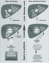
Beneficial effects of stem cells and stem cell-derived factors for liver therapy. Transplantation of hepatocytes derived from stem cells rescues liver diseases by replacing damaged cells and activating host liver regeneration by paracrine manner. Further studies on the paracrine effects revealed that beneficial secretory molecules accelerate the regeneration of host liver and prevent liver fibrosis progression without hepatocyte transplantation. These applications would be a promising therapeutic option for liver disease treatment

References

    1. Corless JK, Middleton HM., 3rd Normal liver function. A basis for understanding hepatic disease. Arch Intern Med. 1983;143:2291–2294. doi: 10.1001/archinte.1983.00350120085018. - DOI - PubMed
    1. Ishibashi H, Nakamura M, Komori A, Migita K, Shimoda S. Liver architecture, cell function, and disease. Semin Immunopathol. 2009;31:399–409. doi: 10.1007/s00281-009-0155-6. - DOI - PubMed
    1. Bernal W, Wendon J. Acute liver failure. N Engl J Med. 2013;369:2525–2534. doi: 10.1056/NEJMra1208937. - DOI - PubMed
    1. Zamora Nava LE, Aguirre Valadez J, Chávez-Tapia NC, Torre A. Acute-on-chronic liver failure: a review. Ther Clin Risk Manag. 2014;10:295–303. - PMC - PubMed
    1. Jadlowiec CC, Taner T. Liver transplantation: current status and challenges. World J Gastroenterol. 2016;22:4438–4445. doi: 10.3748/wjg.v22.i18.4438. - DOI - PMC - PubMed

LinkOut - more resources