. 2017 Sep 27;14(6):775-785.
doi: 10.1007/s13770-017-0081-y. eCollection 2017 Dec.

Potency of Human Urine-Derived Stem Cells for Renal Lineage Differentiation

Affiliations

Potency of Human Urine-Derived Stem Cells for Renal Lineage Differentiation

Jae Young Choi et al. Tissue Eng Regen Med. .

Abstract

Kidney is one of the most difficult organs for regeneration. Several attempts have been performed to regenerate renal tissue using stem cells, the results were not satisfactory. Urine is major product of kidney and contains cells from renal components. Moreover, urine-derived stem cells (USCs) can be easily obtained without any health risks throughout a patient's entire life. Here, we evaluated the utility of USCs for renal tissue regeneration. In this study, the ability of USCs to differentiate into renal lineage cells was compared with that of adipose tissue-derived stem cells (ADSCs) and amniotic fluid-derived stem cells (AFSCs), with respect to surface antigen expression, morphology, immunocytochemistry, renal lineage gene expression, secreted factors, immunomodulatory marker expression, in vivo safety, and renal differentiation potency. Undifferentiated USCs were positive for CD44 and CD73, negative for CD34 and CD45, and formed aggregates after 3 weeks of renal differentiation. Undifferentiated USCs showed high SSEA4 expression, while renal-differentiated cells expressed PAX2, WT1, and CADHERIN 6. In the stem/renal lineage-associated gene analysis, OCT4, SSEA4, and CD117 were significantly downregulated over time, while PAX2, LIM1, PDGFRA, E-CADHERIN, CD24, ACTB, AQP1, OCLN, and NPHS1 were gradually upregulated. In the in vivo safety evaluation, renal-differentiated USCs did not show abnormal histology. These findings demonstrated that USCs have a similar MSC potency, renal lineage-differentiation ability, immunomodulatory effects, and in vivo safety as ADSCs and AFSCs, and showed higher levels of growth factor secretion for paracrine effects. Therefore, urine and USCs can be one of good cell sources for kidney regeneration.

Keywords: Amniotic fluid-derived stem cells; Kidney; Regeneration; Urine-derived stem cells.


Conflict of interest statement

The authors declare that they have no competing interests.All procedures involving animals were performed in accordance with the ethical standards of our institution and following an animal protocol approved by the Yeungnam University Institutional Animal Care and Use Committee (YUMC-AEC2016-003).

Figures

Fig. 1
Fig. 1
Flow cytometric analysis of cell surface marker expression on mesenchymal stem cells. The values were normalized to the isotype IgG control. Expression of these markers was compared to the levels expressed at week 0 (in the undifferentiated phase before induction of renal-lineage differentiation). For urine-derived stem cells (USCs), 95% or more expressed the mesenchymal stem cell markers CD44 and CD73, whereas <3% expressed the hematopoietic lineage markers CD34 and CD45. Ctrl renal stem cells; ADSC adipose tissue-derived stem cells; AFSC amniotic fluid-derived stem cells; USC urine-derived stem cells
Fig. 2
Fig. 2
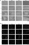
Characterization of in vitro renal lineage-differentiated cells by morphological, immunocytochemical (ICC), quantitative real-time PCR, and secreted trophic factors analysis for 3 weeks. A Representative images from the morphological analysis. The original cell morphology (spindle-like) of the urine-derived stem cells (USCs) gradually changed to a large, round phenotype with a cobble stone-like appearance and cell aggregation. Representative ICC images using SSEA4, Pax2, Wt1, and Cadherin-6 antibodies. B In USCs, the stem cell marker SSEA4 was strongly expressed in undifferentiated stem cells, and then diminished in the differentiated phase. C Pax2 showed widespread nuclear expression at week 0, and then expression gradually became more localized and increased over time, and it was highly expressed in the cell aggregates at week 3. D Wt1 expression in the cytosol gradually increased over time. E Cadherin-6 was expressed in the cytoplasm of a few cells. The target proteins are shown in red, and the nucleus was stained with 4,6–diamidino-2–phenylindole (DAPI, blue). F Real-time PCR analysis. USCs showed the highest expression of LIM1, CD24, and OCLN. G Secreted trophic factor analysis by ELISA. VEGF and PDGF-bb were more strongly expressed in USCs than in ADSCs and AFSCs. The different letters on top of the bars indicate significant differences at p < 0.05. Ctrl renal stem cells, ADSC adipose tissue-derived stem cells, AFSC amniotic fluid-derived stem cells, USC urine-derived stem cells. (Color figure online)
Fig. 2
Fig. 2
Characterization of in vitro renal lineage-differentiated cells by morphological, immunocytochemical (ICC), quantitative real-time PCR, and secreted trophic factors analysis for 3 weeks. A Representative images from the morphological analysis. The original cell morphology (spindle-like) of the urine-derived stem cells (USCs) gradually changed to a large, round phenotype with a cobble stone-like appearance and cell aggregation. Representative ICC images using SSEA4, Pax2, Wt1, and Cadherin-6 antibodies. B In USCs, the stem cell marker SSEA4 was strongly expressed in undifferentiated stem cells, and then diminished in the differentiated phase. C Pax2 showed widespread nuclear expression at week 0, and then expression gradually became more localized and increased over time, and it was highly expressed in the cell aggregates at week 3. D Wt1 expression in the cytosol gradually increased over time. E Cadherin-6 was expressed in the cytoplasm of a few cells. The target proteins are shown in red, and the nucleus was stained with 4,6–diamidino-2–phenylindole (DAPI, blue). F Real-time PCR analysis. USCs showed the highest expression of LIM1, CD24, and OCLN. G Secreted trophic factor analysis by ELISA. VEGF and PDGF-bb were more strongly expressed in USCs than in ADSCs and AFSCs. The different letters on top of the bars indicate significant differences at p < 0.05. Ctrl renal stem cells, ADSC adipose tissue-derived stem cells, AFSC amniotic fluid-derived stem cells, USC urine-derived stem cells. (Color figure online)
Fig. 3
Fig. 3
Flow cytometric analysis of immunomodulatory marker (HLA-DR) expression on MSCs. USCs showed the lowest HLA-DR expression, indicating that USCs do not trigger an immune reaction. HLA-DR, major histocompatibility complex, class II, DR; ADSC adipose tissue-derived stem cells, AFSC amniotic fluid-derived stem cells; USC urine-derived stem cells
Fig. 4
Fig. 4
In vivo safety analysis of renal-differentiated cells. Renal lineage-differentiated USCs were implanted into the subcapsule of the kidney. Four weeks later, a histological analysis was performed. No abnormal morphology was observed in the implanted kidneys. Ctrl renal stem cells, ADSC adipose tissue-derived stem cells, AFSC amniotic fluid-derived stem cells, USC urine-derived stem cells

Similar articles

Cited by

References

    1. Flaquer M, Romagnani P, Cruzado JM. Growth factors and renal regeneration. Nefrologia. 2010;30:385–393. - PubMed
    1. Yokote S, Yokoo T. Organogenesis for kidney regeneration. Curr Opin Organ Transplant. 2013;18:186–190. doi: 10.1097/MOT.0b013e32835f070d. - DOI - PubMed
    1. Hauser PV, De Fazio R, Bruno S, Sdei S, Grange C, Bussolati B, et al. Stem cells derived from human amniotic fluid contribute to acute kidney injury recovery. Am J Pathol. 2010;177:2011–2021. doi: 10.2353/ajpath.2010.091245. - DOI - PMC - PubMed
    1. Breymann C, Schmidt D, Hoerstrup SP. Umbilical cord cells as a source of cardiovascular tissue engineering. Stem Cell Rev. 2006;2:87–92. doi: 10.1007/s12015-006-0014-y. - DOI - PubMed
    1. Graziano A, d’Aquino R, Laino G, Papaccio G. Dental pulp stem cells: a promising tool for bone regeneration. Stem Cell Rev. 2008;4:21–26. doi: 10.1007/s12015-008-9015-3. - DOI - PubMed

LinkOut - more resources