

Calcium Microcrystal Formation in Recurrent Herniation Patients After Autologous Disc Cell Transplantation
- PMID: 30603529
- PMCID: PMC6171663
- DOI: 10.1007/s13770-017-0076-8
Calcium Microcrystal Formation in Recurrent Herniation Patients After Autologous Disc Cell Transplantation
Abstract
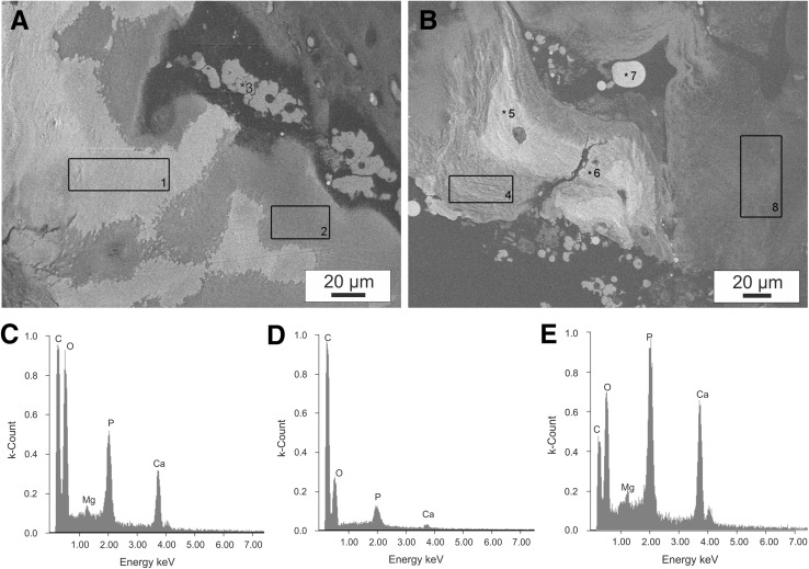
Autologous disc cell transplantation (ADCT) is a cell-based therapy aiming to initiate regeneration of intervertebral disc (IVD) tissue, but little is known about potential risks. This study aims to investigate the presence of structural phenomena accompanying the transformation process after ADCT treatment in IVD disease. Structural phenomena of ADCT-treated patients (Group 1, n = 10) with recurrent disc herniation were compared to conventionally-treated patients with recurrent herniation (Group 2, n = 10) and patients with a first-time herniation (Group 3, n = 10). For ethical reasons, a control group of ADCT patients who did not have a recurrent disc herniation was not possible. Tissue samples were obtained via micro-sequestrectomy after disc herniation and analyzed by micro-computed tomography, scanning electron microscopy, energy dispersive spectroscopy, and histology in terms of calcification zones, tissue structure, cell density, cell morphology, and elemental composition. The major differentiator between sample groups was calcium microcrystal formation in all ADCT samples, not found in any of the control group samples, which may indicate disc degradation. The incorporation of mineral particles provided clear contrast between the different materials and chemical analysis of a single particle indicated the presence of magnesium-containing calcium phosphate. As IVD calcification is a primary indicator of disc degeneration, further investigation of ADCT and detailed investigations assessing each patient's Pfirrmann degeneration grade following herniation is warranted. Structural phenomena unique to ADCT herniation prompt further investigation of the therapy's mechanisms and its effect on IVD tissue. However, the impossibility of a perfect control group limits the generalizable interpretation of the results.
Keywords: Disc calcification; Intervertebral disc; Recurrent disc herniation; Regenerative therapy.
Conflict of interest statement
This work was supported by the German Federal Ministry of Education and Research (BMBF, PtJ-Bio, 0315883) and the DAAD RISE internship program, and the Whitaker Biomedical Engineering Research Fellowship.The study was approved by the independent medical ethical committee as well as the scientific committee from the faculty of medicine Martin-Luther University Halle-Wittenberg and confirmed at 14 July 2010 under approval no. EK-MLU/14072010/hm-bü.
Figures
Similar articles
-
Clinical experience in cell-based therapeutics: disc chondrocyte transplantation A treatment for degenerated or damaged intervertebral disc.Biomol Eng. 2007 Feb;24(1):5-21. doi: 10.1016/j.bioeng.2006.07.002. Epub 2006 Jul 21. Biomol Eng. 2007. PMID: 16963315
-
Clinical experience in cell-based therapeutics: intervention and outcome.Eur Spine J. 2006 Aug;15 Suppl 3(Suppl 3):S397-405. doi: 10.1007/s00586-006-0169-x. Epub 2006 Jul 19. Eur Spine J. 2006. PMID: 16850291 Free PMC article. Clinical Trial.
-
Microstructure analysis method for evaluating degenerated intervertebral disc tissue.Micron. 2017 Jan;92:51-62. doi: 10.1016/j.micron.2016.10.002. Epub 2016 Oct 21. Micron. 2017. PMID: 27871028
-
Intervertebral disc degeneration in the dog. Part 1: Anatomy and physiology of the intervertebral disc and characteristics of intervertebral disc degeneration.Vet J. 2013 Mar;195(3):282-91. doi: 10.1016/j.tvjl.2012.10.024. Epub 2012 Nov 21. Vet J. 2013. PMID: 23177522 Review.
-
[Regenerative medicine of the intervertebral disc: from pathophysiology to clinical application].Med Sci (Paris). 2014 Dec;30(12):1091-100. doi: 10.1051/medsci/20143012012. Epub 2014 Dec 24. Med Sci (Paris). 2014. PMID: 25537039 Review. French.
Cited by
-
In silico modeling the potential clinical effect of growth factor treatment on the metabolism of human nucleus pulposus cells.JOR Spine. 2024 Jul 31;7(3):e1352. doi: 10.1002/jsp2.1352. eCollection 2024 Sep. JOR Spine. 2024. PMID: 39092165 Free PMC article.
-
Comprehensive narrative review on the analysis of outcomes from cell transplantation clinical trials for discogenic low back pain.N Am Spine Soc J. 2022 Dec 22;13:100195. doi: 10.1016/j.xnsj.2022.100195. eCollection 2023 Mar. N Am Spine Soc J. 2022. PMID: 36655116 Free PMC article.
-
Intervertebral Disc Regeneration Injection of a Cell-Loaded Collagen Hydrogel in a Sheep Model.Int J Mol Sci. 2021 Apr 19;22(8):4248. doi: 10.3390/ijms22084248. Int J Mol Sci. 2021. PMID: 33921913 Free PMC article.
-
Evaluation of the Efficacy of Stem Cell Therapy in Animal Models of Intervertebral Disc Degeneration Based on Imaging Indicators: A Systematic Review and Meta-Analysis.Stem Cells Int. 2022 Aug 31;2022:2482653. doi: 10.1155/2022/2482653. eCollection 2022. Stem Cells Int. 2022. PMID: 36093439 Free PMC article.
References
LinkOut - more resources
Full Text Sources
Research Materials