. 2017 Oct 13;15(1):63-73.
doi: 10.1007/s13770-017-0085-7. eCollection 2018 Feb.

Substance-P Ameliorates Dextran Sodium Sulfate-Induced Intestinal Damage by Preserving Tissue Barrier Function

Affiliations

Substance-P Ameliorates Dextran Sodium Sulfate-Induced Intestinal Damage by Preserving Tissue Barrier Function

Dae Yeon Hwang et al. Tissue Eng Regen Med. .

Abstract

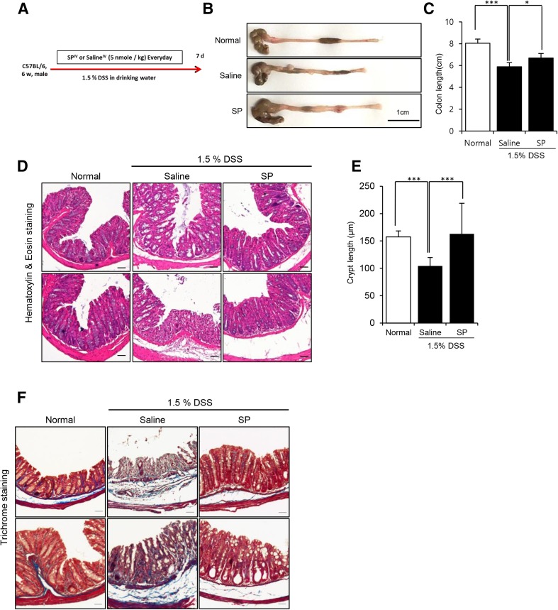
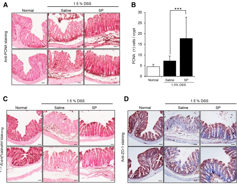
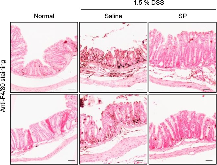
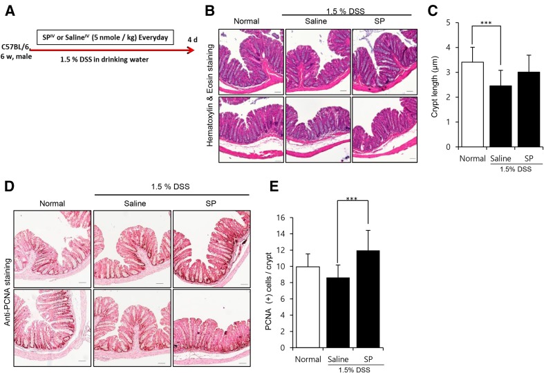
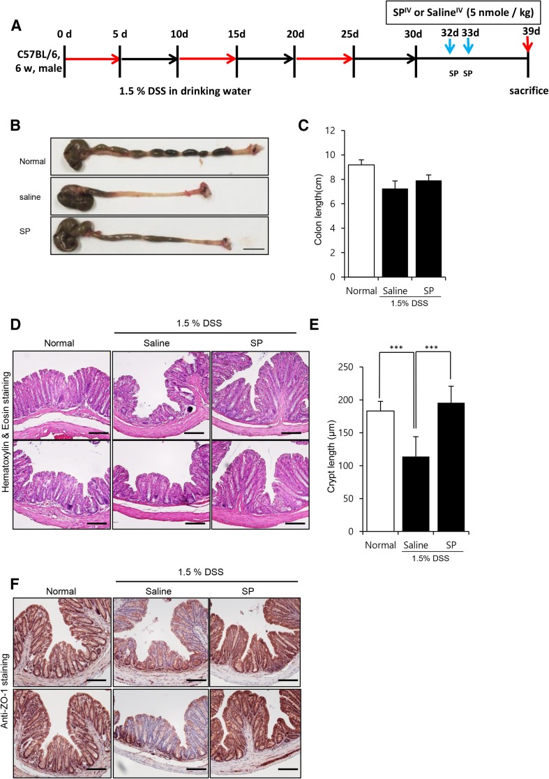
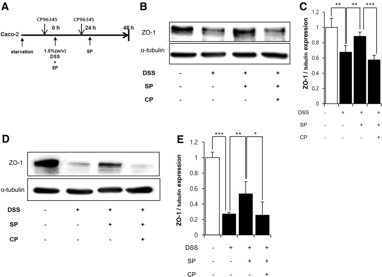
Intestinal inflammation alters immune responses in the mucosa and destroys colon architecture, leading to serious diseases such as inflammatory bowel disease. Thus, the modulation of intestinal integrity and immune responses in IBD can be the critical factor to be considered to reduce the severity of damages. Substance-P (SP), endogenous peptide to be involved in cell proliferation, migration and immune modulation, can exert the therapeutic effect on diverse diseases including cornea damage, rheumatoid arthritis and diabetic complications. SP was found to elevate expression of junctional molecule. Considering the function of SP reported previously, it was inferred that SP is capable of exert the beneficial effect of SP on intestinal diseases by controlling intestinal structure as well as immune responses. In this study, we explored the therapeutic effect of SP on dextran sodium sulfate-induced intestine damage by injecting SP. The effects of SP were evaluated by analyzing crypt structures, proliferating cell pool and infiltration of immune cells. DSS treatment provoked abnormal immune response and disruption of intestine epithelial barrier. However, co-treatment of SP obviously blocked the development of intestinal damages by declining inflammatory responses and sustaining intestinal structure more intact. The treatment of SP to chronic damages also promoted intestinal regeneration by preserving the integrity of colon tissue. Moreover, DSS-induced reduction of epithelial junctional molecule was obviously inhibited by SP treatment in vitro. Taken together, our data indicate that SP can reduce intestinal damages, possibly by modulating barrier structure and immune response. Our results propose SP as candidate therapeutics in intestinal damages.

Keywords: Inflammatory bowel disease; Intestine; Substance-P; Tight junction.


Conflict of interest statement

The authors have no conflict of interest to report.All animal studies were approved by the Ethical Committees for Experimental Animals at Kyung Hee University (Approval number KHMC-IACUC-14-010).

Figures

Fig. 1
Fig. 1
The effect of SP in DSS-induced intestine damages. Mice received the drinking water with 1.5% DSS for 7 days and SP was administrated to tail vein during the intake of DSS. A Experimental schedule for colitis induction and SP treatment. B Gross view of intestine tissue. C Quantification of colon length (cm). D Histological analysis was performed by H&E staining. E Crypt length was measured as a distance from the bottom to apical crypt using Image J program. F Collagen deposition was carried out using Masson’s trichrome staining. Results are shown as mean values ± SD. n = 10/group, *p < 0.05, **p < 0.01, ***p < 0.001. Scale bar: 50 μm
Fig. 2
Fig. 2
The effect of SP on cell repopulation and integrity in DSS-damaged intestine. A PCNA (+) staining to detect the proliferating cells within crypt. B Quantitative analysis in PCNA (+) cells per crypt was carried out. C–D Immunohistochemical staining for E-cadherin and ZO-1. Results are shown as mean values ± SD. n = 10/group, *p < 0.05, **p < 0.01, ***p < 0.001. Scale bar: 50 μm
Fig. 3
Fig. 3
The effect of SP on macrophage infiltration at wounded site. Immunohistochemical staining for f4/80 to check infiltrated macrophage into the injured tissue. Scale bar: 50 μm
Fig. 4
Fig. 4
The effect of SP on DSS-induced intestinal damage at early time post damages. DSS-induced colitis was triggered by exposure to 1.5% DSS. During DSS administration, SP was injected intravenously once a day in regular time and after 4 days, all mice was sacrificed. A Experimental schedule for colitis induction and SP treatment. B H&E staining of damaged tissue from saline- and SP-treated group colonic section. C Crypt length was measured as a distance from the bottom to apical crypt using Image J program. D PCNA staining was carried out to check the effect of SP on proliferation cell pool. E Quantitative analysis in PCN A (+) cells per crypt was performed. Results are shown as mean values ± SD. n = 14/group, *p < 0.05, **p < 0.01, ***p < 0.001. Scale bar: 50 μm
Fig. 5
Fig. 5
The effect of SP on DSS-induced chronic damage of intestines. The chronic colitis was induced by administering DSS repeatedly and then, SP was injected to tail vein of mice for two consecutive days. A Experimental schedule for colitis induction and SP treatment. B Gross view of intestine tissue from each group. C Colon length was quantified (cm) D H&E staining of damaged tissue from saline- and SP-treated group colonic section. E Crypt length was measured as a distance from the bottom to apical crypt using Image J program. F Immunohistochemical staining for ZO-1 expression. Results are shown as mean values ± SD. n = 10/group, *p < 0.05, **p < 0.01, ***p < 0.001. Scale bar: 100 μm
Fig. 6
Fig. 6
The effect of SP on ZO-1 expression in DSS-damaged intestinal epithelial cell in vitro. Caco-2 was pretreated with NK-1R antagonist (CP96345) for 30 min, followed by 1.5% DSS (w/v) and SP (100 nM) exposure for 24 or 48 h. 24 h later, SP treatment was repeated. A The experimental scheme to CP96345 and SP treatment. B–C At 24 h post DSS treatment, ZO-1 expression level was determined by western blots. ZO-1 expression in Caco-2 was quantified relatively. D–E At 48 h post DSS treatment, ZO-1 expression level was determined by western blots. ZO-1 expression in Caco-2 was quantified relatively. Results were shown as mean values ± SD. All experiments were performed in triplicate. The data was expressed as the means of three independent experiments. CP: CP 96345, NK-1R antagonist

Similar articles

Cited by

References

    1. Zois CD, Katsanos KH, Kosmidou M, Tsianos EV. Neurologic manifestations in inflammatory bowel diseases: current knowledge and novel insights. J Crohns Colitis. 2010;4:115–124. doi: 10.1016/j.crohns.2009.10.005. - DOI - PubMed
    1. Sun X, Yamada H, Shibata K, Muta H, Tani K, Podack ER, et al. CD30 ligand is a target for a novel biological therapy against colitis associated with Th17 responses. J Immunol. 2010;185:7671–7680. doi: 10.4049/jimmunol.1002229. - DOI - PubMed
    1. Zhang J, Fu S, Sun S, Li Z, Guo B. Inflammasome activation has an important role in the development of spontaneous colitis. Mucosal Immunol. 2014;7:1139–1150. doi: 10.1038/mi.2014.1. - DOI - PMC - PubMed
    1. Kaistha A, Levine J. Inflammatory bowel disease: the classic gastrointestinal autoimmune disease. Curr Probl Pediatr Adolesc Health Care. 2014;44:328–334. doi: 10.1016/j.cppeds.2014.10.003. - DOI - PubMed
    1. Landy J, Ronde E, English N, Clark SK, Hart AL, Knight SC, et al. Tight junctions in inflammatory bowel diseases and inflammatory bowel disease associated colorectal cancer. World J Gastroenterol. 2016;22:3117–3126. doi: 10.3748/wjg.v22.i11.3117. - DOI - PMC - PubMed

LinkOut - more resources