. 2018 Feb 3;15(3):275-285.
doi: 10.1007/s13770-018-0113-2. eCollection 2018 Jun.

In Vivo Observation of Endothelial Cell-Assisted Vascularization in Pancreatic Cancer Xenograft Engineering

Affiliations

In Vivo Observation of Endothelial Cell-Assisted Vascularization in Pancreatic Cancer Xenograft Engineering

Boyoung Jung et al. Tissue Eng Regen Med. .

Abstract

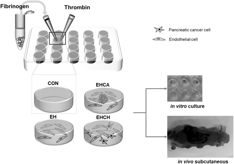
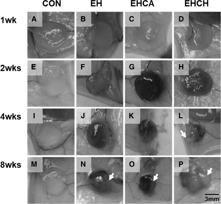
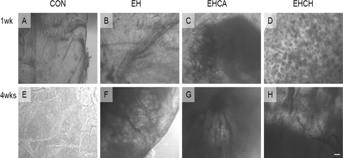
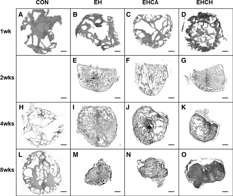
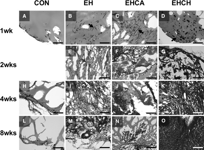
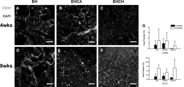
In this study, for better understanding of patient-derived xenograft (PDX) generation, angiogenic characteristics during PDX cancerous tissue generation was investigated with different initial cell seeding conditions in the hydrogel. We monitored the angiogenic changes during the formation of in vivo cancer cell line xenografts induced by endothelial cells. Our in vivo cancer tissue formation system was designed with the assistance of tissue engineering technology to mimic patient-derived xenograft formation. Endothelial cells and MIA PaCa-2 pancreatic carcinoma cells were encapsulated in fibrin gel at different mixing configurations and subcutaneously implanted into nude mice. To investigate the effect of the initial cancerous cell distribution in the fibrin gel, MIA PaCa-2 cells were encapsulated as a homogeneous cell distribution or as a cell aggregate, with endothelial cells homogeneously distributed in the fibrin gel. Histological observation of the explanted tissues after different implantation periods revealed three different stages: isolated vascular tubes, leaky blood vessels, and mature cancerous tissue formation. The in vivo engineered cancerous tissues had leaky blood vessels with low expression of the vascular tight junction marker CD31. Under our experimental conditions, complex cancer-like tissue formation was most successful when tumorous cells and endothelial cells were homogeneously mixed in the fibrin gel. The present study implies that tumorous xenograft tissue formation can be achieved with a low number of initial cells and that effective vascularization conditions can be attained with a limited volume of patient-derived cancer tissue. Endothelial cell-assisted vascularization can be a potent choice for the effective development of vascularized cancerous tissues for studying patient-derived xenografts, cancer angiogenesis, cancer metastasis, and anticancer drugs.

Keywords: Cancer tissue engineering; Pancreatic cancer; Patient-derived xenograft; Vascularization.


Conflict of interest statement

There are no conflicts to declare.The animal experiment protocol was reviewed and approved by the institutional animal care and use committee of Asan Medical Center (Protocol Number: 2014-12-003).

Figures

Fig. 1
Fig. 1
Schematic diagram of the pancreatic cancer xenograft engineering. Four experimental groups were prepared according to embedded cell and sample conditions: (1) control with no cells in the fibrin (CON), (2) endothelial cells homogeneously distributed in the fibrin (EH), (3) endothelial cells homogeneously distributed with the cancer aggregate located in the center of the fibrin (EHCA), (4) endothelial cells and cancer cells homogeneously distributed in the fibrin (EHCH)
Fig. 2
Fig. 2
Gross observation of in vivo tissue explants. At 1, 2, 4, and 8 weeks of after implantation, the samples were removed and observed. A, E, I, M Control group, B, F, J, N EH group, C, G, K, O EHCA group and D, H, L, P EHCH group
Fig. 3
Fig. 3
Optical microscope observation of the harvested samples after 1 and 4-week implantation. A, E Control group, B, F EH group, C, G EHCA group and D, H EHCH group. Scale bar = 200 μm
Fig. 4
Fig. 4
Gross observation of in vivo implanted fibrin gel after H&E staining. Images are adjusted to same scale for visual comparison of samples size. A, H, L Control group, B, E, I, M EH group, C, F, J, N EHCA group and D, G, K, O EHCH group. Scale bar = 500 μm
Fig. 5
Fig. 5
Histological observation of in vivo implanted cell embedded fibrin gel. At 1, 2, 4, and 8 weeks of after implantation, the samples were H&E staining. A, H, L Control group, B, E, I, M EH group, C, F, J, N EHCA group and D, G, K, O EHCH group. Scale bar = 200 μm. (Color figure online)
Fig. 6
Fig. 6
Blood vessel length comparison after 1-week implantation. A EH group, B EHCA group, C EHCH group and D cross-group comparison graph
Fig. 7
Fig. 7
Immunofluorescence image of vascular marker von Wilbrand factor (vWF, green) expression in in vivo implanted fibrin gels for 4- and 8-week. All endothelial cell mixed explants were stained with vWF implementing blood vessel formation. A, D EH group, B, E EHCA group and C, F EHCH group. Scale bar = 100 μm. (Color figure online)
Fig. 8
Fig. 8
Vasculogenesis in the in vivo pancreatic cancer xenograft tissue model. Immunofluorescence image of CD31 (green) and α-SMA (red) expression in in vivo implanted fibrin gels for 4- and 8-week. A, D EH group, B, E EHCA group and C, F EHCH group. Scale bar = 50 μm. G Area fraction of alpha smooth muscle actin and CD31 expression. (Color figure online)

Similar articles

Cited by

References

    1. Bianco P, Robey PG. Stem cells in tissue engineering. Nature. 2001;414:118–121. doi: 10.1038/35102181. - DOI - PubMed
    1. Langer R, Vacanti JP. Tissue engineering. Science. 1993;260:920–926. doi: 10.1126/science.8493529. - DOI - PubMed
    1. Karam JP, Muscari C, Montero-Menei CN. Combining adult stem cells and polymeric devices for tissue engineering in infarcted myocardium. Biomaterials. 2012;33:5683–5695. doi: 10.1016/j.biomaterials.2012.04.028. - DOI - PubMed
    1. Zhang K, Huang D, Yan Z, Wang C. Heparin/collagen encapsulating nerve growth factor multilayers coated aligned PLLA nanofibrous scaffolds for nerve tissue engineering. J Biomed Mater Res A. 2017;105:1900–1910. doi: 10.1002/jbm.a.36053. - DOI - PubMed
    1. Hoganson DM, Bassett EK, Vacanti JP. Lung tissue engineering. Front Biosci (Landmark Ed). 2014;19:1227–1239. doi: 10.2741/4278. - DOI - PubMed

LinkOut - more resources