

Positive Effects of Bisphosphonates on Osteogenic Differentiation in Patient-Derived Mesenchymal Stem Cells for the Treatment of Osteoporosis
- PMID: 30603570
- PMCID: PMC6171649
- DOI: 10.1007/s13770-018-0127-9
Positive Effects of Bisphosphonates on Osteogenic Differentiation in Patient-Derived Mesenchymal Stem Cells for the Treatment of Osteoporosis
Abstract
Background: Recent evidence from in vitro and in vivo studies indicates that bisphosphonates may promote osteoblastic bone formation and potently inhibit osteoclast activity. However, little is known about the potential effect of bisphosphonates on the recruitment of osteoblastic precursors from patient-derived bone marrow stromal cells due to difficulties in accessing human bone marrow from healthy and disease subjects.
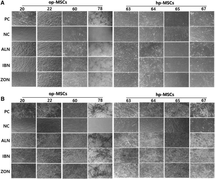
Methods: In this study, we evaluated the potential of using FDA-approved and clinically utilized bisphosphonates such as alendronate, ibandronate, and zoledronate to enhance the development of bone forming osteoblasts from osteoporosis patient- and healthy-person derived hBMSCs (op-MSCs and hp-MSCs, respectively). hBMSCs were obtained from postmenopausal women without endocrine diseases or receiving hormone replacement therapy. Cells were treated with or without a bisphosphonate (alendronate, ibandronate, and zoledronate) and analyzed over 21 days of culture.
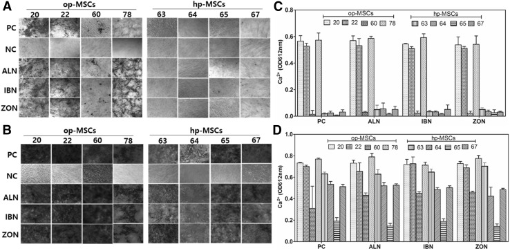
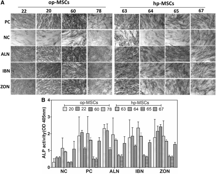
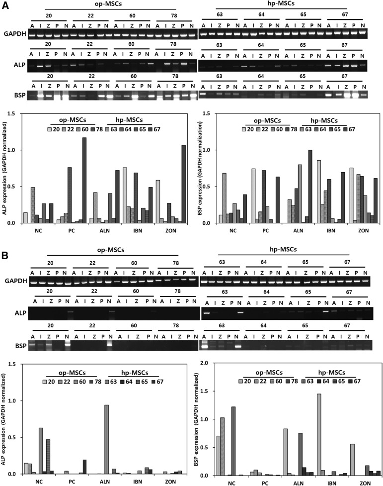
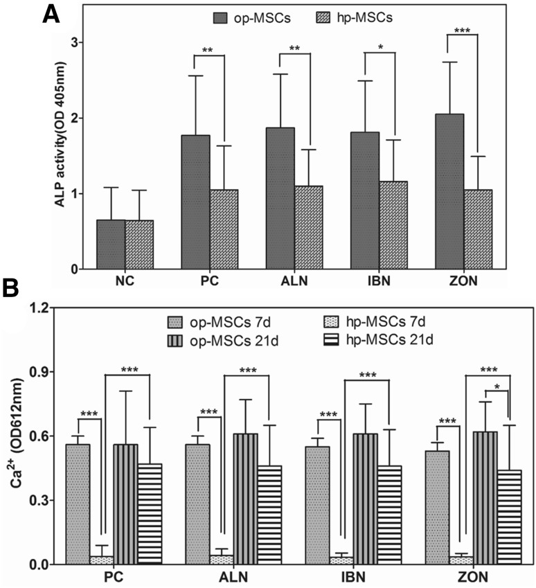
Results: hBMSC from osteoporosis-patient with bisphosphonates treatment demonstrated a significant increase in Alizarin red staining after 7 days compared to that from healthy-person. Calcium contents and alkaline phosphatase (ALP) enzyme activity also demonstrated an increased propensity in hMSCs from osteoporosis-patient compared to those from healthy-person, although there were inter-individual variations. Gene expression levels varied among different donors. There were no significant differences in the effect on the osteoblastic differentiation of hBMSCs among alendronate, ibandronate, and zoledronate. Statistical significance in the osteoblastic differentiation of hBMSCs between the positive control group cultured in osteogenic medium alone and groups cultured in osteogenic medium supplemented with bisphosphonate was not shown either. These results might be due to various cell types of hBMSCs from individual clinical patients and concentrations of bisphosphonate used.
Conclusion: Our study using a clinically relevant in vitro model suggests that bisphosphonate treatment is more effective for patients with osteoporosis than its preventive effect for healthy person. In addition, patient-specific responses to bisphosphonates should be considered rather than bisphosphonate type prior to prescription. Further investigations are needed to determine how bisphosphonates influence hBMSCs function to mediate bone quality and turnover in osteoporotic patients. Such studies can generate novel approaches to treat age-related osteoporotic bone loss.
Keywords: Bisphosphonates; Mesenchymal stem cell; Osteoblast; Osteoporosis.
Conflict of interest statement
The authors declare that they have no conflict of interest.Institutional review board approval and patient consenting was obtained for this study (IRB No. 06-2010-142).
Figures
References
LinkOut - more resources
Full Text Sources
Research Materials
Miscellaneous