Review
. 2018 Aug 27;15(5):547-557.
doi: 10.1007/s13770-018-0155-5. eCollection 2018 Oct.

In Situ Cross-Linkable Hydrogels as a Dynamic Matrix for Tissue Regenerative Medicine

Affiliations
Review

In Situ Cross-Linkable Hydrogels as a Dynamic Matrix for Tissue Regenerative Medicine

Kyung Min Park et al. Tissue Eng Regen Med. .

Abstract

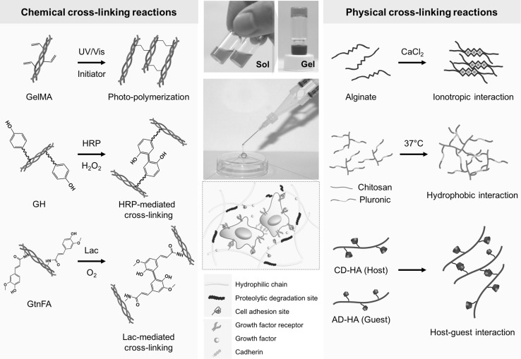
Background: Polymeric hydrogels are extensively used as promising biomaterials in a broad range of biomedical applications, including tissue engineering, regenerative medicine, and drug delivery. These materials have advantages such as structural similarity to the native extracellular matrix (ECM), multi-tunable physicochemical and biological properties, and biocompatibility.

Methods: In situ forming hydrogels show a phase transition from a solution to a gel state through various physical and chemical cross-linking reactions. These advanced hydrogel materials have been widely used for tissue regenerative medicine because of the ease of encapsulating therapeutic agents, such as cells, drugs, proteins, and genes.

Results: With advances in biomaterials engineering, these hydrogel materials have been utilized as either artificial cellular microenvironments to create engineered tissue constructs or as bioactive acellular matrices to stimulate the native ECM for enhanced tissue regeneration and restoration.

Conclusion: In this review, we discuss the use of in situ cross-linkable hydrogels in tissue engineering and regenerative medicine applications. In particular, we focus on emerging technologies as a powerful therapeutic tool for tissue regenerative medicine applications.

Keywords: In situ cross-linkable hydrogels; Polymeric hydrogels; Tissue engineering; Tissue regenerative medicine.


Conflict of interest statement

The authors declare that they have no competing interests.There are no animal experiments carried out for this article.

Figures

Fig. 1
Fig. 1
Cross-linking strategies to fabricate in situ forming hydrogels. In situ cross-linkable hydrogels exhibit phase transition from solution to gel state through physical and chemical cross-linking reactions. These advanced hydrogel materials can serve as dynamic cellular or acellular matrices for tissue engineering and regenerative medicine. Abbreviations: GelMA, gelatin-methacryloyl; GH, gelatin-g-hydroxyphenyl propionic acid; HRP, horseradish peroxidase; GtnFA, gelatin-g-ferulic acid; Lac, laccase; CD-HA, b-cyclodextrin-conjugated hyaluronic acid; AD-HA; adamantine-modified hyaluronic acid (adapted with permission from Ref. [2, 3])
Fig. 2
Fig. 2
Engineered vascular constructs using photo-curable GelMA hydrogels. A Schematic representation of the stepwise process of endothelial lumen formation in the engineered microenvironment. B Premature vessel formation of ECFCs surrounded by α-smooth muscle actin (α-SMA)-expressing MSCs (yellow arrow). Scale bar is 20 µm. C Functional vascular formation in vivo. Immunohistochemistry exhibited that the engineered vasculatures were positively stained for human CD31 (red arrow) and murine capillaries (green arrow), carrying murine erythrocytes (asterisks). Fluorescence images show sections stained with rhodamine-conjugated UEA-1 lectin (to mark human ECFC-lined vessels) and fluorescein isothiocyanate-conjugated anti-α-SMA (to mark perivascular cells; red arrowheads). UEA-1 lectin did not bind to the murine vessels (green arrow). Scale bars are 10 µm and 50 µm (adapted with permission from Ref [53]). (Color figure online)
Fig. 3
Fig. 3
Hypoxia-inducible hydrogels to create engineered vasculatures. A Schematic diagram of vascular morphogenesis of ECFCs within artificial hypoxic microenvironment. B Confocal microscopic images of ECFCs cultured within non-hypoxic gels (NG) and hypoxic gels (HG); confocal Z-stacks and orthogonal sections exhibited lumen formation (yellow arrow). Scale bar is 50 µm. C Quantification of vascular tube formation (mean tube coverage, tube length, and tube thickness). D Real-time reverse-transcription polymerase chain reaction for gene expression of ECFCs cultured within two types of hydrogels (NG vs. HG), which is relevant to vascular morphogenesis. Results in C and D are shown as the average value ± s.d. Significance levels were set as *p < 0.05, **p < 0.01, and ***p < 0.001 (adapted with permission from Ref. [16]). (Color figure online)
Fig. 4
Fig. 4
H2O2-controllable hydrogels for facilitating neovascularization though transient upregulation of intracellular ROS levels in ECs. A Schematic representation of in situ hydrogel formation via dual enzyme-mediated cross-linking reaction. Newly formed covalent bonds are indicated in red. Transient upregulation of intracellular ROS levels of ECs by sustained release of H2O2 from hydrogels. B Representative optical and fluorescent microscopic images. Scale bar is 100 μm. C Quantitative analysis of fluorescence-positive cells. The results in B are shown as an average ± s.d. Significance levels were set at *p < 0.05, **p < 0.01, and ***p < 0.001. ## indicates not significant. D In ovo angiogenic effect of the hydrogels. Histological sections of hydrogels seven days after injection, stained with α-SMA. Scale bar is 100 μm (adapted with permission from Ref. [17]). (Color figure online)
Fig. 5
Fig. 5
Chemokine-loaded in situ forming GH hydrogels for enhanced wound healing in diabetic mouse models. A Schematic illustration of in situ GH hydrogel formation through HRP-mediated cross-linking reaction, encapsulating cell-recruiting chemokines (IL-8 or MIP-3α). B Representative digital images depicting wound healing by the chemokine-loaded GH hydrogels in streptozotocin-induced diabetic mice on day 0, 7, 14, and 21. C Re-epithelialization of the regenerative tissues on day 7 after hydrogel treatment. The explants were subjected to Masson’s trichrome staining and showed re-epithelialization of the damaged tissues with the hydrogels. The arrows indicate regenerated edges of the skin wound. D dermis; E epidermis; EG epithelial gap; GT granulation tissue (adapted with permission from Ref. [73])

Similar articles

Cited by

References

    1. Zhang Yu Shrike, Khademhosseini Ali. Advances in engineering hydrogels. Science. 2017;356(6337):eaaf3627. doi: 10.1126/science.aaf3627. - DOI - PMC - PubMed
    1. Park KM, Lewis D, Gerecht S. Bioinspired hydrogels to engineer cancer microenvironments. Annu Rev Biomed Eng. 2017;19:109–133. doi: 10.1146/annurev-bioeng-071516-044619. - DOI - PMC - PubMed
    1. Park S, Park KM. Engineered polymeric hydrogels for 3D tissue models. Polymers. 2016;8:23. doi: 10.3390/polym8010023. - DOI - PMC - PubMed
    1. Hoffman AS. Hydrogels for biomedical applications. Adv Drug Deliv Rev. 2012;64:18–23. doi: 10.1016/j.addr.2012.09.010. - DOI - PubMed
    1. Mahapatra C, Jin GZ, Kim HW. Alginate-hyaluronic acid-collagen composite hydrogel favorable for the culture of chondrocytes and their phenotype maintenance. Tissue Eng Regen Med. 2016;13:538–546. doi: 10.1007/s13770-016-0059-1. - DOI - PMC - PubMed

LinkOut - more resources