Review
. 2018 Jul 25;15(6):673-697.
doi: 10.1007/s13770-018-0135-9. eCollection 2018 Dec.

Collagen Scaffolds in Cartilage Tissue Engineering and Relevant Approaches for Future Development

Affiliations
Review

Collagen Scaffolds in Cartilage Tissue Engineering and Relevant Approaches for Future Development

Vincent Irawan et al. Tissue Eng Regen Med. .

Abstract

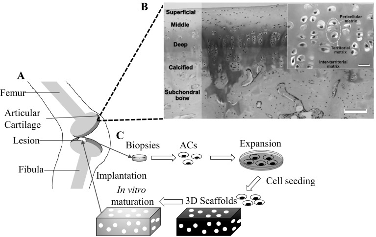
Background: Cartilage tissue engineering (CTE) aims to obtain a structure mimicking native cartilage tissue through the combination of relevant cells, three-dimensional scaffolds, and extraneous signals. Implantation of 'matured' constructs is thus expected to provide solution for treating large injury of articular cartilage. Type I collagen is widely used as scaffolds for CTE products undergoing clinical trial, owing to its ubiquitous biocompatibility and vast clinical approval. However, the long-term performance of pure type I collagen scaffolds would suffer from its limited chondrogenic capacity and inferior mechanical properties. This paper aims to provide insights necessary for advancing type I collagen scaffolds in the CTE applications.

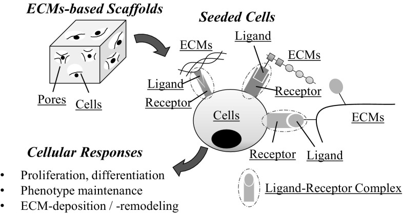
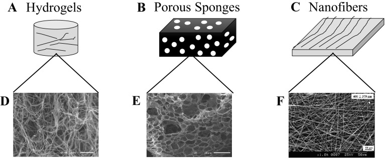
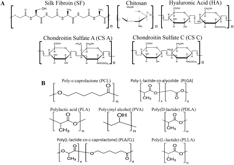
Methods: Initially, the interactions of type I/II collagen with CTE-relevant cells [i.e., articular chondrocytes (ACs) and mesenchymal stem cells (MSCs)] are discussed. Next, the physical features and chemical composition of the scaffolds crucial to support chondrogenic activities of AC and MSC are highlighted. Attempts to optimize the collagen scaffolds by blending with natural/synthetic polymers are described. Hybrid strategy in which collagen and structural polymers are combined in non-blending manner is detailed.

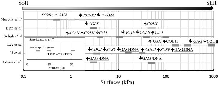
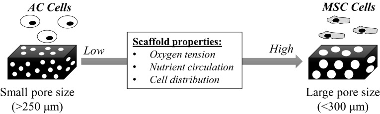
Results: Type I collagen is sufficient to support cellular activities of ACs and MSCs; however it shows limited chondrogenic performance than type II collagen. Nonetheless, type I collagen is the clinically feasible option since type II collagen shows arthritogenic potency. Physical features of scaffolds such as internal structure, pore size, stiffness, etc. are shown to be crucial in influencing the differentiation fate and secreting extracellular matrixes from ACs and MSCs. Collagen can be blended with native or synthetic polymer to improve the mechanical and bioactivities of final composites. However, the versatility of blending strategy is limited due to denaturation of type I collagen at harsh processing condition. Hybrid strategy is successful in maximizing bioactivity of collagen scaffolds and mechanical robustness of structural polymer.

Conclusion: Considering the previous improvements of physical and compositional properties of collagen scaffolds and recent manufacturing developments of structural polymer, it is concluded that hybrid strategy is a promising approach to advance further collagen-based scaffolds in CTE.

Keywords: Articular chondrocytes; Cartilage tissue engineering; Hybrid scaffolds; Mesenchymal stem cells; Type I collagen.


Conflict of interest statement

The authors have no financial conflict of interest.There are no human experiments conducted for this article and approval of institutional review board (IRB) is not necessary for this article. There are no animal experiments carried out for this article.

Figures

Fig. 1
Fig. 1
Illustration of articular cartilage tissues and concept of cartilage tissue engineering. A Schematic representation of articular cartilage with a lesion in knee. B Articular cartilage consists of multiple zones and is separated with subchondral bone by calcified zones. Some areas are stained with red Safranin O/Fast staining, indicating presence of proteoglycan. Inset shown AC (dotted circle) embedded in cartilage matrix. Scale bar: 20 μm. C Flow of cartilage tissue engineering started with biopsy of cartilage tissue and ended with implantation of mature scaffolds into lesions. (Parts of figure B adapted with permission from Armiento et al. [6])
Fig. 2
Fig. 2
Schematic illustration of the attachment of cells to ECMs-based scaffolds. Cells attach to the ECMs molecules by formation of ligand-receptor complex. Cells express various receptors capable of recognizing various type of ligand presented by ECMs molecules. Activated cell receptor subsequently modify various responses of cells (proliferation, ECMs deposition)
Fig. 3
Fig. 3
Schematic illustration of macroscopic and internal structures of collagen A, B hydrogels, C, D sponges, and E, F nanofibers. (F was reprinted with permission from Yeo et al. [123]. Copyright (2008) American Chemical Society)
Fig. 4
Fig. 4
Effect of substrate stiffness (colored boxes) on gene transcription (italic name; Sox9, α-SMA, COL II, Col I, Col X, ACAN) and protein synthesize (underlined name; GAG/DNA, COLII) of ACs (blue and red colored) and MSCs (green and yellow colored). Blue and red boxes respectively indicate positive and negative changes of differentiated phenotype of ACs, as reported by Sanz-Ramoz et al. [94], Schuh et al. [26, 95], Li et al. [27] and Lee et al. [83]. In a similar manner, green and yellow boxes are associated with changes of chondrogenic markers of MSCs as reported by Bian et al. [37] and Murphy et al. [31]. (Color figure online)
Fig. 5
Fig. 5
Schematic illustration of change of scaffold properties with evolution of pore or mesh sizes. Small pore sized scaffolds support the differentiated phenotype of ACs due to non-even cell distribution (induce cell aggregation) and low oxygen tension. On the other hand, large pore sized scaffolds enhance the chondrogenic differentiation of MSCs due to higher oxygen tension and better nutrition circulation
Fig. 6
Fig. 6
Chemical structures of A natural polymers [silk fibroin (SF), chitosan, hyaluronic acid (HA), chondroitin sulfate-A (CS-A), chondroitin-6 sulfate (CS-C)] commonly blended with collagen matrices (sponges, hydrogel, nanofibers) and B synthetic polymers [poly(lactic acid) (PLA), poly(L-lactide) (PLLA), poly(ε-caprolactone) (PCL), poly(vinyl alcohol) (PVA), poly(D-lactide) (PDLA), poly(L-lactide-co-e-caprolactone) (PLA/CL), poly(L-lactideco-glycolide) (PLGA)] incorporated with collagen matrices in non-blended manner
Fig. 7
Fig. 7
Structure and properties of collagen/silk fibroin (Col/SF) scaffolds. A Microstructure of freeze-dried SF scaffold. B Microstructure of freeze-dried Col/SF scaffolds with 20 wt% Col and 4 wt% SF. C Microstructure of Col/SF scaffolds incorporated with TGF-β1 containing poly(lactic-co-glycolic-acid) microsphere (TGF-β1- COL/SF). D Histological evaluation of Col/SF scaffolds after implantation in vivo for 12 weeks. COL/SF and no scaffold as a control group. (Parts of figure are adapted with the permission from [128] A, B and [125] C and D)
Fig. 8
Fig. 8
Structure and properties of collagen/chitosan scaffolds. A Microstructure of freeze-dried collagen/chitosan scaffold. B Change of mechanical properties of collagen/chitosan scaffold caused by the variation of chitosan concentration (0–50 wt%). C Remaining of collagen/chitosan scaffold after incubation for 28 days in serum containing media. Data (n = 3) is plotted in mean ± standard deviation. Significance is indicated by *p < 0.05; **p < 0.01; ***p < 0.001. D, E Determination of sGAG deposition of collagen (collagen:chitosan at 100:0) versus collagen/chitosan (collagen:chitosan at 75:25) scaffolds, by Safranin O staining (D) and sGAG quantification (E). Data (n = 3) is plotted in mean ± standard deviation. Significance is indicated by *p < 0.05; **p < 0.01; ***p < 0.001. (Parts of figure are adapted, with permission, from [137] A, B and [139] C, D, E)
Fig. 9
Fig. 9
A, B Histological sections of scaffolds of collagen (A) and collagen/HA (B). Red color indicates sGAG staining by Safranin-O. C Effect of HA concentration of the collagen/HA scaffolds on the total amount of deposited chondroitin sulfate. Data (n = 6) is plotted in mean ± standard deviation. *indicates insignificant differences. † indicates p < 0.01. (Parts of figure are adapted from [35] A, B and [144] C). (Color figure online)
Fig. 10
Fig. 10
A Sox9 expression of MSCs seeded in scaffold of collagen/chondroitin sulfate (CCS) with different compressive modulus (stiffness). B Amount of CS (chondroitin sulfate) in two different type of collagen scaffolds at initial time and after 3 days of incubation in cell culture media. 2af-CS and 2rf CS indicate afibrillar type II collagen-CS and fibrillar type II collagen-CS scaffolds, respectively. (Parts of figure are adapted from [31] A with permission and [34] B under a Creative Commons Attribution License)
Fig. 11
Fig. 11
A Conceptual illustration of hybrid scaffolds in which it is composed of two different components: housing framework and bioactive filler. BD are the examples of collagen-based hybrid scaffolds. B PLGA mesh (left figure) was furnished with collagen web to form hybrid scaffold (right figure). C 3D printed PLGA (left figure) was used as housing framework for collagen sponges to obtain hybrid scaffold (right figure). D Appearance of hybrid scaffold of nanofibers type I/II collagen (left figure) and the corresponding internal structure (right figure). (Parts of figure are adapted, with permission, from [177] B, [178] C and [179] D)
Fig. 12
Fig. 12
Hybrid scaffold of collagen PLGA mesh/collagen web. A Initial microstructure of PLGA mesh/collagen web, B, C microstructure of PLGA mesh/collagen web after 1 and 4 weeks of chondrocyte incorporation. Chondrocyte was attached and suspended on the collagen web. P and C indicate PLGA mesh and collagen web, respectively. D Northern blot analyses of chondrocytes seeded in PLGA mesh/collagen web scaffolds for the gene encoding ColI, COLII, and ACAN for the period of 0, 2, 4, 12 weeks. (Parts of figure are adapted from [180] A, B, C, and D with permission)
Fig. 13
Fig. 13
Hybrid scaffolds of type II collagen/PLGA obtained by fused deposition manufacturing (FDM). A Illustration of PLGA scaffolds obtained by stacking four layers of extruded polymer fiber in different angle (4D:0°, 45°, 90°, 135°). dh and Φn indicate fiber distance and nozzle aperture, respectively. B Cell number of chondrocytes and C deposited amount of GAG by chondrocytes seeded on hybrid scaffold of different parameters. Sample naming is 4D/dh and 8D/dh. FD is freeze-dried type II collagen. n = 3, p < 0.05, * and # are significantly higher and lower from other scaffolds. (Parts of figure are adapted from [178] A, B, C with permission)
Fig. 14
Fig. 14
A Hybrid scaffolds of PLLA obtained by fused-deposition method (FDM) with different aperture size (1, 1.5, 2 mm) and collagen. Control is a collagen gel. B Number of macrophages accumulated in the hybrid scaffolds of collagen and various synthetic polymer (PLLA, PLGA(L), PLGA(H), PLA/CL, and PDLA after 2 months of subcutaneous implantations in mice. (Parts of figure are adapted from [182] with permission)

References

    1. Mankin HJ. The response of articular cartilage to mechanical injury. J Bone Joint Surg Am. 1982;64:460–466. - PubMed
    1. Pearle AD, Warren RF, Rodeo SA. Basic science of articular cartilage and osteoarthritis. Clin Sports Med. 2005;24:1–12. - PubMed
    1. Hunziker EB. Articular cartilage repair: basic science and clinical progress. A review of the current status and prospects. Osteoarthritis Cartilage. 2002;10:432–463. - PubMed
    1. O’Driscoll SW. The healing and regeneration of articular cartilage. J Bone Joint Surg Am. 1998;80:1795–1812. - PubMed
    1. Camp CL, Stuart MJ, Krych AJ. Current concepts of articular cartilage restoration techniques in the knee. Sports Health. 2014;6:265–273. - PMC - PubMed

LinkOut - more resources