Case Reports
. 2018 Dec;6(23):463.
doi: 10.21037/atm.2018.11.13.

Downhill esophageal varices: a therapeutic dilemma

Affiliations
Case Reports

Downhill esophageal varices: a therapeutic dilemma

Raja Chandra Chakinala et al. Ann Transl Med. 2018 Dec.

Abstract

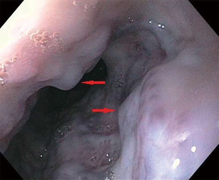
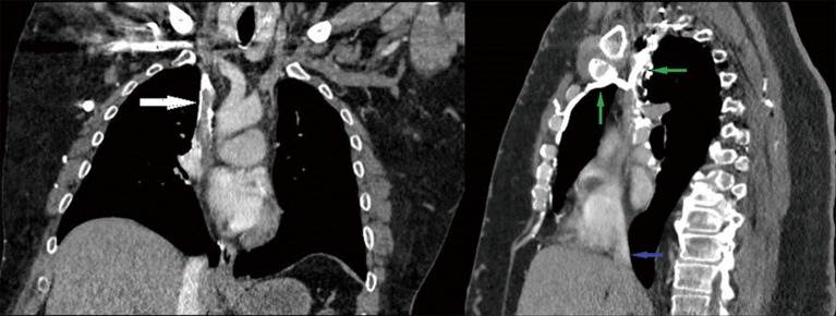
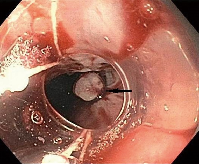
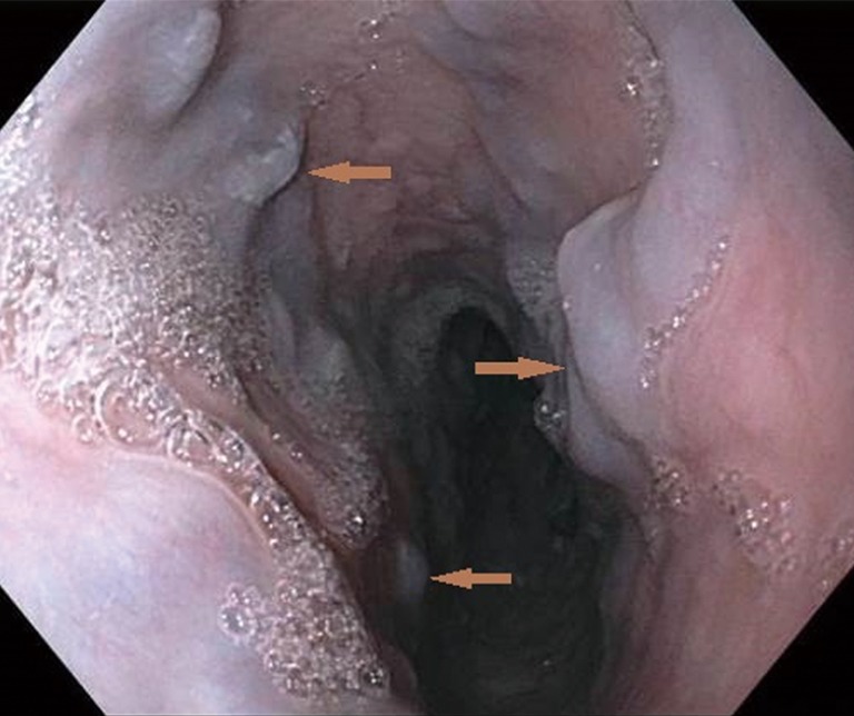
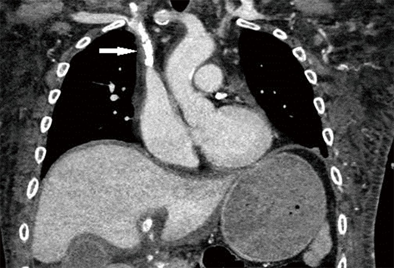
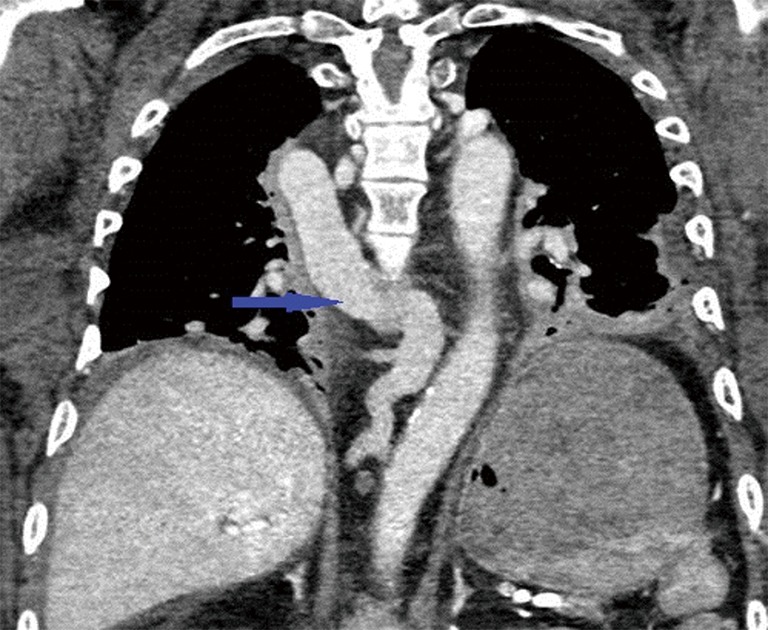
Esophageal varices can cause life-threatening complications and are most often a sequela of liver disease. Although a rare cause of gastrointestinal bleeding, downhill variceal bleeding secondary to superior vena cava (SVC) obstruction should be considered in the differential diagnosis for patients with upper gastrointestinal hemorrhage. We discuss two such cases of downhill esophageal varices presenting with hematemesis in patients with end stage renal disease and no history of cirrhosis. These varices were thought to be secondary to SVC occlusion caused by complications from previous dialysis catheters. However, their difficult anatomy posed a significant challenge to the therapeutic interventions.

Keywords: Downhill esophageal varices; gastrointestinal bleeding; superior vena cava obstruction (SVC obstruction).


Conflict of interest statement

Conflicts of Interest: The authors have no conflicts of interest to declare.

Figures

Figure 1
Figure 1
Upper endoscopy image showing large varices in in the proximal and middle third of the esophagus (red arrows).
Figure 2
Figure 2
CT coronal section demonstrating occlusion of the superior vena cava (white arrow). CT sagittal section showing extensive collaterals (green arrows) and patent inferior vena cava (blue arrow).
Figure 3
Figure 3
Band ligation of a varix (black arrow) was performed on the repeat upper endoscopy.
Figure 4
Figure 4
Upper endoscopy image showing medium-sized varices in the proximal and mid esophagus (brown arrows). No stigmata of bleeding was visualized.
Figure 5
Figure 5
CT delayed contrast image demonstrating occlusion of the cranial superior vena cava (white arrow).
Figure 6
Figure 6
Prominent azygous vein (blue arrow) noticed in the mediastinum on coronal section of CT angiogram of the thorax.

Similar articles

Cited by

References

    1. Areia M, Romãozinho JM, Ferreira M, et al. "Downhill" varices. A rare cause of esophageal hemorrhage. Rev Esp Enferm Dig 2006;98:359-61. 10.4321/S1130-01082006000500006 - DOI - PubMed
    1. Harwani YP, Kumar A, Chaudhary A, et al. Combined uphill and downhill varices as a consequence of rheumatic heart disease: a unique presentation. J Clin Exp Hepatol 2014;4:63-5. 10.1016/j.jceh.2013.10.003 - DOI - PMC - PubMed
    1. Jammal GE. An unusual case of Esophageal Varices: “Downhill” type. Gastroenterol Hepatol Open Access 2015;2:00040.
    1. Rhoades DP, Forde KA, Tabibian JH. Proximal Esophageal Varices: A Rare Yet Treatable Cause of Hemorrhage. Clin Gastroenterol Hepatol 2016;14:e105-6. 10.1016/j.cgh.2016.03.029 - DOI - PubMed
    1. Sheiner NM, Palayew MJ. "Downhill" esophageal varices in superior vena caval obstruction. Can Med Assoc J 1969;100:961-4. - PMC - PubMed

Publication types