Review
. 2019 Jan 17;11(3):799-819.
doi: 10.1039/c8nr07769j.

Applications of nanoparticles in biomedical imaging

Affiliations
Review

Applications of nanoparticles in biomedical imaging

Xiangjun Han et al. Nanoscale. .

Abstract

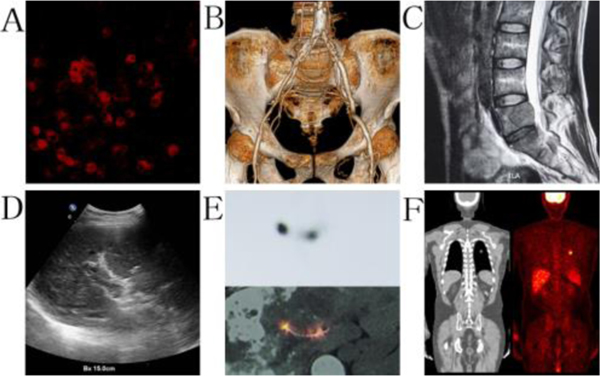
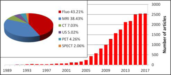
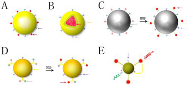
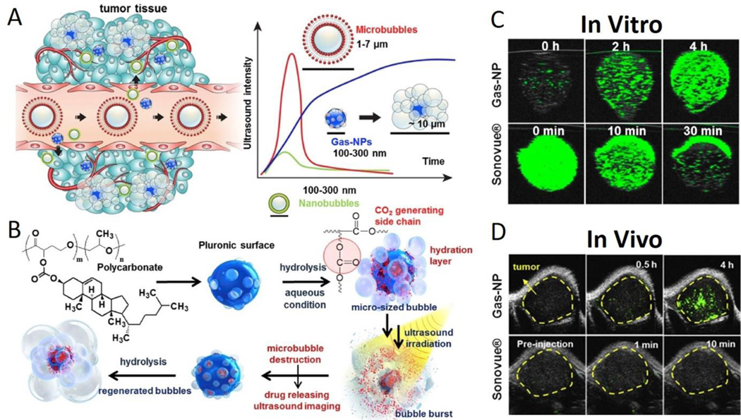
An urgent need for early detection and diagnosis of diseases continuously pushes the advancements of imaging modalities and contrast agents. Current challenges remain for fast and detailed imaging of tissue microstructures and lesion characterization that could be achieved via development of nontoxic contrast agents with longer circulation time. Nanoparticle technology offers this possibility. Here, we review nanoparticle-based contrast agents employed in most common biomedical imaging modalities, including fluorescence imaging, MRI, CT, US, PET and SPECT, addressing their structure related features, advantages and limitations. Furthermore, their applications in each imaging modality are also reviewed using commonly studied examples. Future research will investigate multifunctional nanoplatforms to address safety, efficacy and theranostic capabilities. Nanoparticles as imaging contrast agents have promise to greatly benefit clinical practice.


Conflict of interest statement

Conflicts of interest

There are no conflicts to declare.

Figures

Fig. 1
Fig. 1
The primary imaging technologies in biomedical practice, including A: fluorescence image of tumor cells; B: CT diagnosis for artery stenosis; C: MRI image of lumber cancer metastasis; D: US detection of portal vein thrombosis; E: SPECT evaluation for 125I seeds implantation; and F: PET detection of lung cancer tumor. All images were obtained from medical imaging research institute of China Medical University.
Fig. 2
Fig. 2
The number of publications searching for “nanoparticle and imaging” in Pubmed is rapidly increasing each year. Fluorescence and MRI imaging modalities represent the greatest areas of activity.
Fig. 3
Fig. 3
Different structure and composition of nanoparticles in fluorescence biomedical imaging. A: vector type; B: core-shell structure of NP; C: NP as a quencher; D: NP connected with fluorophore and quencher, E: Forster resonance energy transferred imaging NP (yellow arrow: nanoparticle; red arrow: fluorescent dye; blue arrow: ligand; grey arrow: quencher; yellow curve arrow: energy transfer; green curve arrow: excitation light; red curve arrow: emission light; MMP: matrix metalloproteinase).
Fig. 4
Fig. 4
A safer T1 MRI contrast in a compact plasmonic nanoparticle with enhanced fluorescence was synthesized by a multilayer core-shell nanostructure, and known as nanomatryoshka (NM). Each NM consisted of an Au core, an Au shell and silica spacing layer encapsulated magnetic metal and fluorescent dye. This form protected fluorescent dye and reduced the metal release. Fe (III)-NM exhibited a 2× greater relaxivity than current MRI contrast agent (Gd-DOTA), and the photostability of fluorescent dye significantly increased (23×). Lower panel evaluated MRI and fluorescence imaging of FeCy7-NM in vivo. (A) Untreated in MRI, (B) Treated in MRI, red circle is nanoparticle and blue circle is saline, (C) Fluorescence imaging after injection. This nanoparticle can enable not only powerful tissue visualization with MRI but also fluorescence-based nanoparticle tracking. Reprinted with permission. Copyright 2018. American Chemical Society.
Fig. 5
Fig. 5
Different structures of MRI/CT imaging nanoparticles. A: basic structure; B core-shell structure; C: vector structure; D: mixed structure; E: core-shell structure. (Black arrow: MRI/CT imaging materials; Blue arrow: surface decoration.
Fig. 6
Fig. 6
A CT-based molecular imaging nanoparticle was developed as blood pool and myocardial scar specific imaging contrast agent. Management of patients suffering from myocardial infarction is based on the extent of coronary artery stenosis and myocardial scar burden. (Upper panel) Gold nanoparticles (AuNPs) functionalized with collagen-binding adhesion protein 35 (CNA35) play the vascular imaging role at early phase and molecular imaging at late phase. (Lower panel) Animal experiment demonstrated a specific myocardial infaction imaging at 6 hours after injection (A). Control rat with myocardial infarction after injection of AuNPs (B) and rat without myocardial infaction after injection of CNA35-AuNPs (C) did not generate specific CT enhancement. This nanoparticle demonstrates a potential use for coronary artery imaging and myocardial infarction evaluation. Reproduced with permission. Copyright 2018. Elsevier.
Fig. 7
Fig. 7
(A) Gas-NPs (blue nanoparticles) were synthesized via the O/W emulsion method with a size of 290nm and accumulated in tumor through the EPR effect. Current ultrasound contrast agents and large-sized perfluorocarbons-encapsulated microbubbles (red microparticles) present strong echo signals, but the large dimension prevents extravasation from vessel to surrounding tumor tissue. Also, small-sized nanobubbles (green nanoparticles) demonstrate a good biological distribution and effective extravasation, but the echo signals are not strong enough. (B) Chemical structure of hydrolysable carbonate copolymer; Poly(cholesteryl butyrolacone-co-propylene oxide). The carbonate copolymer was emulsified to produce solidified Gas-NPs via the O/W emulsion method. The Gas-NPs start to hydrolyze to produce CO2 nanobubbles in aqueous condition, followed by expansion/coalescence of nanobubbles into microbubbles for the targeted tumor US imaging. In addition, the anticancer drug-loaded Gas-NPs enable US-triggered drug delivery. (C) The US imaging test in vitro demonstrated a gradually CO2 generating process, current ultrasound contrast agent (Sonovue®) was the control (D) The ultrasound imaging ability in vivo showed a strong and persisted signals in whole tumor, current ultrasound contrast agent (Sonovue®) was the control. This nanoparticle demonstrates the unique and beneficial chemical gas-generating mechanism and is potentially useful for real-time ultrasound imaging and cancer therapy. Reproduced with permission. Copyright 2016. Elsevier.
Fig. 8
Fig. 8
A PET imaging nanoparticle was synthesized from PVPh polymer and labeled with 124I or 125I, and then coated with specific targeting antibodies. Which endows a satisfactory endothelium PET imaging (lung). Reproduced with permission. Copyright 2012. Elsevier.
Fig. 9
Fig. 9
A dual-modality nanoparticle was developed with Ag2Se quantum dots for fluorescence imaging and Gd-DTPA for MRI imaging. The excellent imaging efficiency indicate the potential value for multimodal imaging in clinical and scientific applications. Reproduced with permission. Copyright 2018. Royal Society of Chemistry.
Fig. 10
Fig. 10
A theranostic nanoplatform was synthesized from thiol-capped Bi nanoparticles. They have a high X-ray attenuation coefficient and a strong photothermal conversion efficiency, which endow them with the simultaneous CT imaging, radiotherapy and thermotherapy. Reproduced with permission. Copyright 2018. Elsevier.

Similar articles

Cited by

References

    1. Jung DH, Hwang S, Song GW, Ahn CS, Moon DB, Ha TY, Kim KH, Park GC, Kim BS, Park IJ, Lim SB, Kim JC, Yoo MW, Byeon JS, Jung HY, Lee GH, Myung SJ, Choe J, Choi JY, Park HW and Lee SG, Transplant Proc, 2016, 48, 145–151. - PubMed
    1. Lu WL, Jansen L, Post WJ, Bonnema J, Van de Velde JC and De Bock GH, Breast Cancer Res Treat, 2009, 114, 403–412. - PubMed
    1. Ryvolova M, Chomoucka J, Drbohlavova J, Kopel P, Babula P, Hynek D, Adam V, Eckschlager T, Hubalek J, Stiborova M, Kaiser J and Kizek R, Sensors (Basel), 2012, 12, 14792–14820. - PMC - PubMed
    1. Torres Martin de Rosales R, Tavare R, Glaria A, Varma G, Protti A and Blower PJ, Bioconjug Chem, 2011, 22, 455–465. - PMC - PubMed
    1. Fan Q, Cheng K, Hu X, Ma X, Zhang R, Yang M, Lu X, Xing L, Huang W, Gambhir SS and Cheng Z, J Am Chem Soc, 2014, 136, 15185–15194. - PMC - PubMed