. 2019 Feb;71(1):329-344.
doi: 10.1007/s10616-018-0289-2. Epub 2019 Jan 2.

A serum-free medium suitable for maintaining cell morphology and liver-specific function in induced human hepatocytes

Affiliations

A serum-free medium suitable for maintaining cell morphology and liver-specific function in induced human hepatocytes

Pan-Pan Li et al. Cytotechnology. 2019 Feb.

Abstract

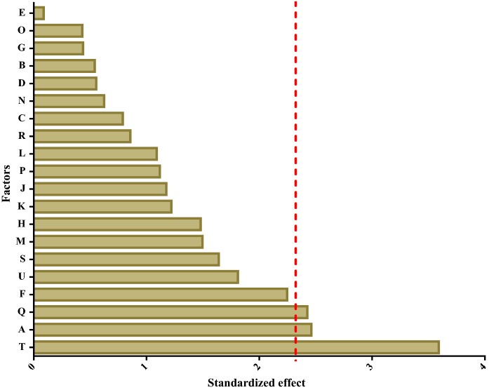
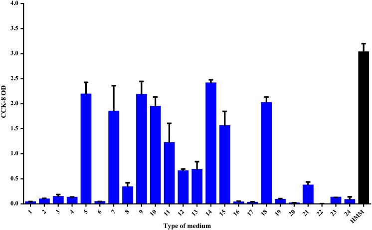
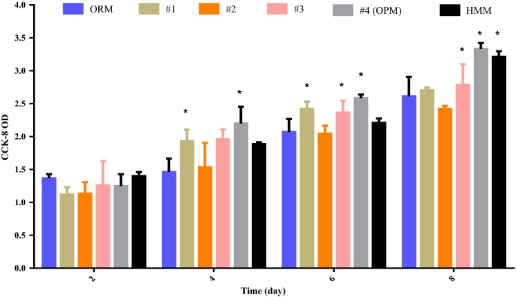
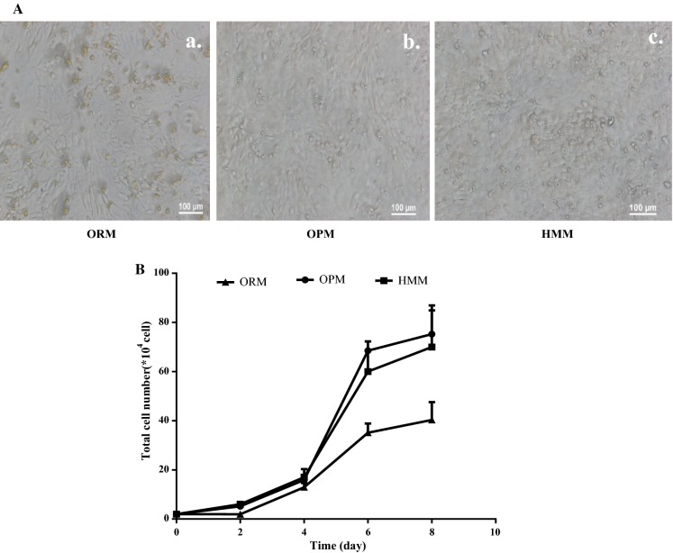
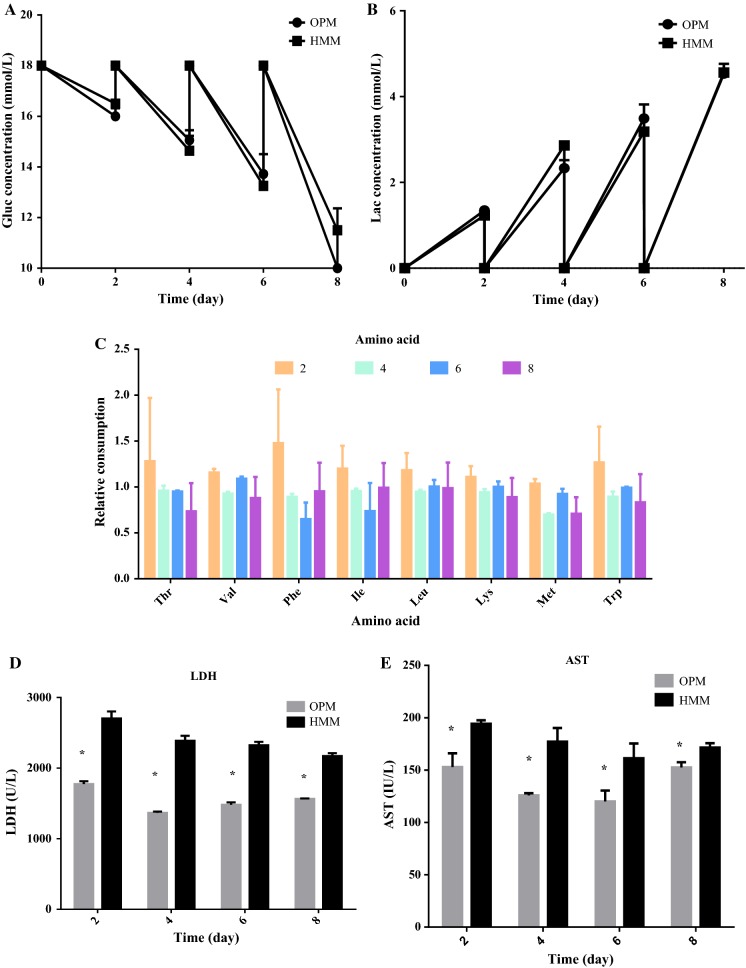
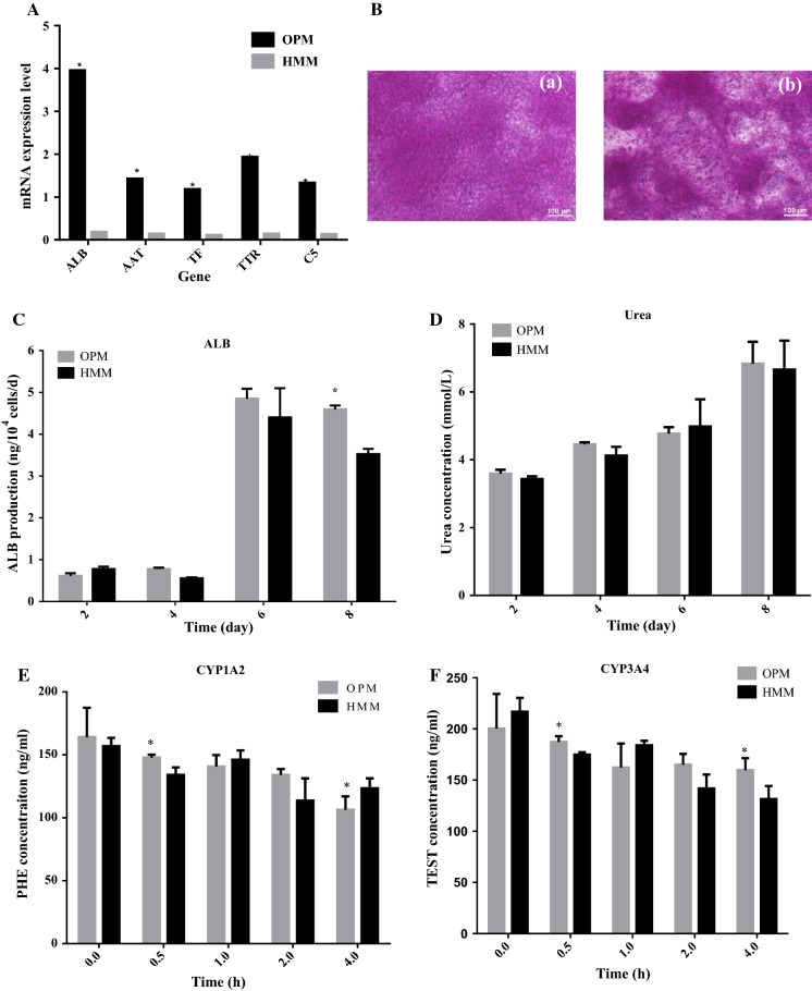
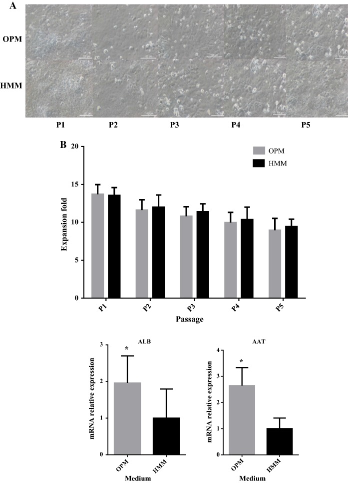
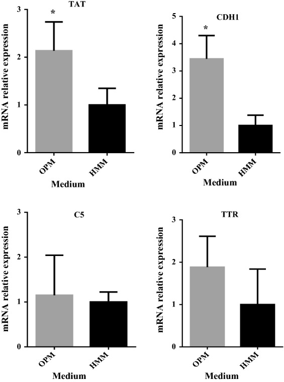
hiHep is a new type of hepatocyte-like cell that is predicted to be a potential unlimited source of hepatocytes for a bioartificial liver. However, hiHep cannot currently be used in clinical settings because serum must be added during the culture process. Thus, a defined medium is required. Because serum is complex, an efficient statistical approach based on the Plackett-Burman design was used. In this manner, an original medium and several significant cell growth factors were identified. These factors include insulin, VH, and VE, and the original medium was optimized based on these significant factors. Additionally, hiHep liver-specific functions and metabolism in the optimized serum-free medium were measured. Results showed that hiHep functions, such as glycogen storage, albumin secretion, and urea production, were well maintained in our optimized serum-free medium. In summary, we created a chemically defined, serum-free medium in which cell growth, proliferation, metabolism, and function were well maintained. This medium has the potential to support the clinical use of hiHep.

Keywords: Bioartificial liver; Medium; Serum-free; hiHep.


Figures

Fig. 1
Fig. 1
The effect of factors in the medium. Normal plot of the standardized effects of cell growth factors in a Plackett–Burman design (The significance level of each variable effect was determined using t distribution associated with Student’s t-test,; The red vertical line means the t critical value (α = 0.1). (Color figure online)
Fig. 2
Fig. 2
Screening of the original medium. hiHep was seeded in 24-well plates and cultured in the indicated medium for 8 days. After culturing, cells were subjected to CCK-8 analysis to evaluate their growth. Cells cultured in HMM served as the control
Fig. 3
Fig. 3
The effect of significant factors on cell growth. a The growth of hiHep cells in different media that were supplemented with respective factors. 1: ORM + insulin, 2: ORM + VE, 3: ORM + VH, 4: ORM + VE + VH + insulin (medium 4 was called the optimized medium). Data are present as the mean ± SD of six independent experiments. *p < 0.05 versus ORM
Fig. 4
Fig. 4
Cell growth results. A hiHep morphology in ORM, OPM and HMM; (a) ORM; (b)OPM; (c) HMM. B The hiHep growth curves
Fig. 5
Fig. 5
Characterization of hiHep metabolism and damage. a Glucose concentration; b Lactate production; c relative consumption of eight essential amino acids (The variation of amino acids concentration in OPM versus the corresponding of HMM); d LDH activity; e Transaminase activity. Data are presented as the mean ± SD of three independent experiments. *p < 0.05
Fig. 6
Fig. 6
Assessment of hiHep cell functionality. A Liver-specific gene analysis by semi-quantitative PCR; B Cells on day 8 showed the capability to store glycogen, as seen in the areas stained in pink. (a) OPM, (b) HMM; C Albumin secretion during the culture process; D Urea production during the culture process; E CYP1A2 activity; F CYP3A4 activity (The activity of CYP was reflected by the reduction of corresponding substrate concentration, the greater variation means the higher activity). *p < 0.05 versus HMM
Fig. 7
Fig. 7
Long-term cell culture. a hiHep cell morphology in each passage (100 μΜ); b hiHep cell proliferation in each passage; c mRNA expression levels in liver-specific functions (i.e., ALB, AAT, TAT, CDH1, C5and TTR) in hiHep cells as tested by qRT-PCR. The data are expressed as the mean ± SD (n = 3). *p < 0.05 versus HMM
Fig. 7
Fig. 7
Long-term cell culture. a hiHep cell morphology in each passage (100 μΜ); b hiHep cell proliferation in each passage; c mRNA expression levels in liver-specific functions (i.e., ALB, AAT, TAT, CDH1, C5and TTR) in hiHep cells as tested by qRT-PCR. The data are expressed as the mean ± SD (n = 3). *p < 0.05 versus HMM

Similar articles

Cited by

References

    1. Aravindan V, Gnanaraj J, Madhavi S, Liu HK. Lithium-ion conducting electrolyte salts for lithium batteries. Chemistry. 2011;17:14326–14346. doi: 10.1002/chem.201101486. - DOI - PubMed
    1. Butt H, Mehmood A, Ali M, Tasneem S, Anjum MS, Tarar MN, Khan SN, Riazuddin S. Protective role of vitamin E preconditioning of human dermal fibroblasts against thermal stress in vitro. Life Sci. 2017;184:1–9. doi: 10.1016/j.lfs.2017.07.002. - DOI - PubMed
    1. Carpentier B, Gautier A, Legallais C. Artificial and bioartificial liver devices: present and future. Gut. 2009;58:1690–1702. doi: 10.1136/gut.2008.175380. - DOI - PubMed
    1. Ekpenyong MG, Antai SP, Asitok AD, Ekpo BO. Plackett–Burman design and response surface optimization of medium trace nutrients for glycolipopeptide biosurfactant production. Iran Biomed J. 2017;21:249–260. doi: 10.18869/acadpub.ibj.21.4.249. - DOI - PMC - PubMed
    1. Guillouzo A, Corlu A, Aninat C, Glaise D, Morel F, Guguen-Guillouzo C. The human hepatoma HepaRG cells: a highly differentiated model for studies of liver metabolism and toxicity of xenobiotics. Chem Biol Interact. 2007;168:66–73. doi: 10.1016/j.cbi.2006.12.003. - DOI - PubMed

LinkOut - more resources