

Establishment of renal proximal tubule cell lines derived from the kidney of p53 knockout mice
- PMID: 30603921
- PMCID: PMC6368522
- DOI: 10.1007/s10616-018-0261-1
Establishment of renal proximal tubule cell lines derived from the kidney of p53 knockout mice
Abstract
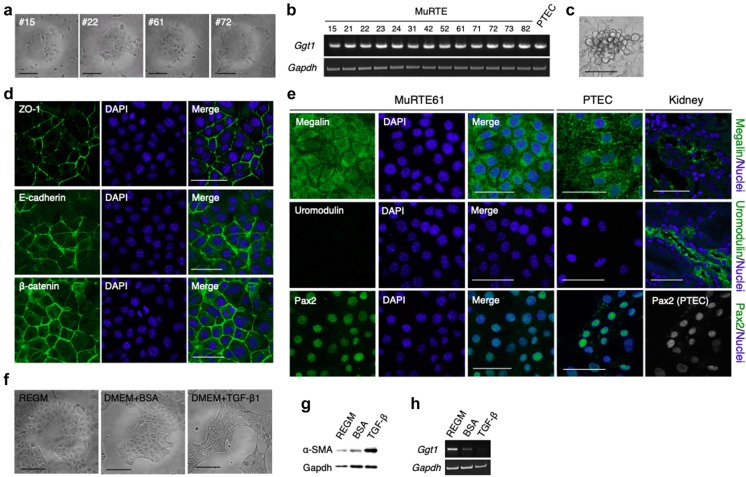
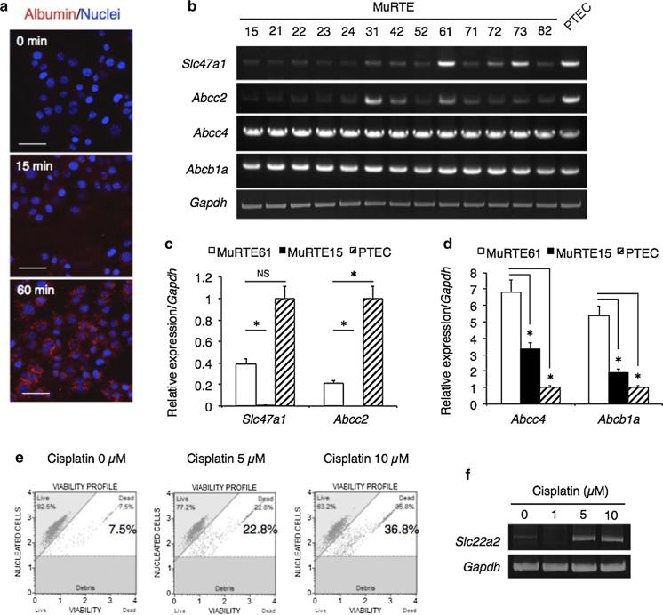
The human cell line HK-2 is most commonly used as a model of renal proximal tubular epithelial cells (PTECs) for various studies despite the absence or low expression of transporters characteristic of parental PTECs. In an effort to develop reliable PTEC models, several human cell lines have been newly established over the last decade. In contrast, reliable mouse PTEC models are still unavailable. In this study, we established immortalized renal cortex tubule cell lines derived from p53 knockout mice and evaluated various PTEC characteristics toward the development of reliable mouse PTEC models. Here, we focus on MuRTE61, one of 13 newly established clonal cell lines. Albumin uptake in MuRTE61 cells was verified by incubation with fluorescent dye-labeled albumin. RT-PCR confirmed the expression of efflux transporter genes characteristic of PTECs in the MuRTE61 cells. MuRTE61 cells exhibited high sensitivity to treatment with cisplatin, a nephrotoxic agent, accompanied by upregulated expression of the uptake transporter Slc22a2 gene. Furthermore, MuRTE61 cells consistently formed spheroids with a lumen and apicobasal polarity in three-dimensional Matrigel cultures. Apical brush border microvilli were also observed in the spheroids by transmission electron microscopy. These data validate that MuRTE61 can be characterized as a reliable mouse PTEC line. In future, detailed analysis of reliable mouse and human PTEC lines will provide an accurate extrapolation of results of experiments using mice and humans, and may help resolve apparent inconsistencies between mouse and human nephrotoxicity.
Keywords: 3D cell culture; Cell line; Cell spheroids; Matrigel; Renal proximal tubular epithelial cells.
Conflict of interest statement
Conflict of interest
The authors declare no conflict interest.
Human and animal rights
All research was conducted according to the Regulation for the Care and Use of Laboratory Animals of Kitasato University. Animal experimentation protocol was approved by President of Kitasato University through the judgment by Institutional Animal Care and Use Committee of Kitasato University (Approval ID: No. 15-168).
Figures

Similar articles
-
A Human Proximal Tubular Epithelial Cell Model to Explore a Knowledge Gap on Neonatal Drug Disposition.Curr Pharm Des. 2017;23(38):5911-5918. doi: 10.2174/1381612823666171009143146. Curr Pharm Des. 2017. PMID: 28990525
-
Functional differentiation and scalable production of renal proximal tubular epithelial cells from human pluripotent stem cells in a dynamic culture system.Cell Prolif. 2022 Mar;55(3):e13190. doi: 10.1111/cpr.13190. Epub 2022 Jan 31. Cell Prolif. 2022. PMID: 35102634 Free PMC article.
-
Flow stimulates drug transport in a human kidney proximal tubule-on-a-chip independent of primary cilia.Biochim Biophys Acta Gen Subj. 2020 Jan;1864(1):129433. doi: 10.1016/j.bbagen.2019.129433. Epub 2019 Sep 11. Biochim Biophys Acta Gen Subj. 2020. PMID: 31520681
-
Current State of In vitro Cell-Based Renal Models.Curr Drug Metab. 2018;19(4):310-326. doi: 10.2174/1389200219666180119115133. Curr Drug Metab. 2018. PMID: 29357789 Review.
-
Renal proximal tubule cell cultures for studying drug-induced nephrotoxicity and modulation of phenotype expression by medium components.Ren Fail. 1992;14(3):371-83. doi: 10.3109/08860229209106645. Ren Fail. 1992. PMID: 1509170 Review.
Cited by
-
Delivery and Transcriptome Assessment of an In Vitro Three-Dimensional Proximal Tubule Model Established by Human Kidney 2 Cells in Clinical Gelatin Sponges.Int J Mol Sci. 2023 Oct 24;24(21):15547. doi: 10.3390/ijms242115547. Int J Mol Sci. 2023. PMID: 37958530 Free PMC article.
-
A Novel Protocol for Culturing Polarized Proximal Tubular Epithelial Cells from Kidney Biopsies: Enhancing Platforms for Drug Excretion and Nephrotoxicity Studies.J Xenobiot. 2025 Apr 1;15(2):52. doi: 10.3390/jox15020052. J Xenobiot. 2025. PMID: 40278157 Free PMC article.
-
Amphiphilic nebramine analogs synergize with β-lactam/β-lactamase inhibitor combinations, including cefepime-taniborbactam and meropenem-xeruborbactam against metallo-β-lactamase-carrying Pseudomonas aeruginosa.RSC Med Chem. 2025 Jul 17. doi: 10.1039/d5md00375j. Online ahead of print. RSC Med Chem. 2025. PMID: 40734664 Free PMC article.
-
DNA damage in proximal tubules triggers systemic metabolic dysfunction through epigenetically altered macrophages.Nat Commun. 2025 Apr 28;16(1):3958. doi: 10.1038/s41467-025-59297-x. Nat Commun. 2025. PMID: 40295524 Free PMC article.
-
Insights into Repeated Renal Injury Using RNA-Seq with Two New RPTEC Cell Lines.Int J Mol Sci. 2023 Sep 18;24(18):14228. doi: 10.3390/ijms241814228. Int J Mol Sci. 2023. PMID: 37762531 Free PMC article.
References
Grants and funding
LinkOut - more resources
Full Text Sources
Molecular Biology Databases
Research Materials
Miscellaneous
