. 2019 Mar;137(3):487-500.
doi: 10.1007/s00401-018-1946-4. Epub 2019 Jan 2.

C9orf72 arginine-rich dipeptide proteins interact with ribosomal proteins in vivo to induce a toxic translational arrest that is rescued by eIF1A

Affiliations

C9orf72 arginine-rich dipeptide proteins interact with ribosomal proteins in vivo to induce a toxic translational arrest that is rescued by eIF1A

Thomas G Moens et al. Acta Neuropathol. 2019 Mar.

Abstract

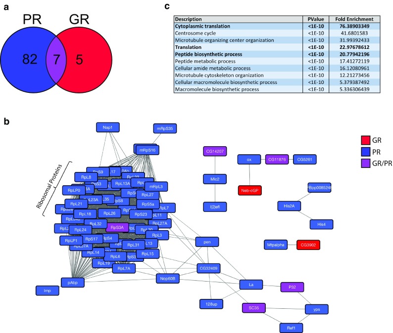
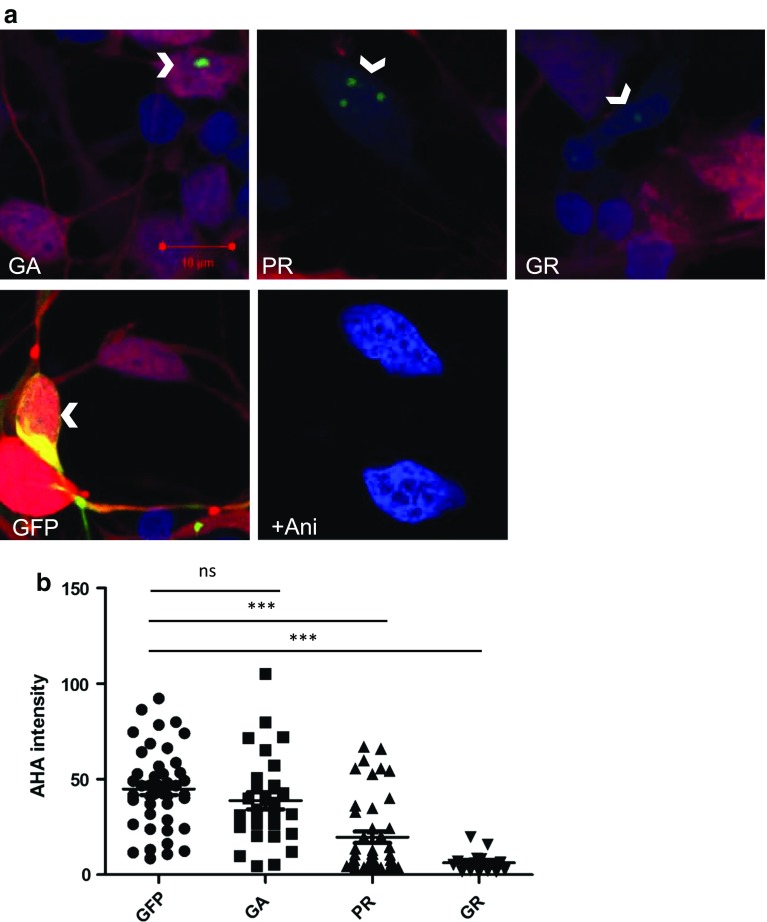
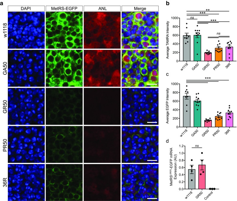
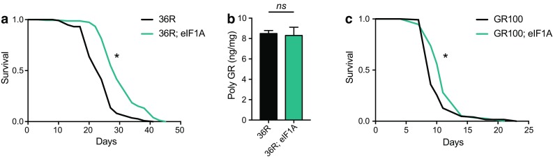
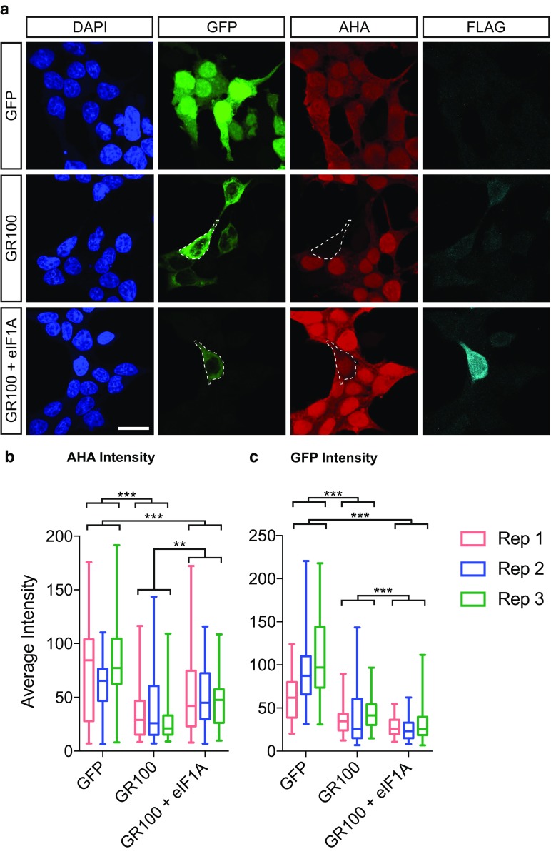
A GGGGCC hexanucleotide repeat expansion within the C9orf72 gene is the most common genetic cause of both amyotrophic lateral sclerosis and frontotemporal dementia. Sense and antisense repeat-containing transcripts undergo repeat-associated non-AUG-initiated translation to produce five dipeptide proteins (DPRs). The polyGR and polyPR DPRs are extremely toxic when expressed in Drosophila neurons. To determine the mechanism that mediates this toxicity, we purified DPRs from the Drosophila brain and used mass spectrometry to identify the in vivo neuronal DPR interactome. PolyGR and polyPR interact with ribosomal proteins, and inhibit translation in both human iPSC-derived motor neurons, and adult Drosophila neurons. We next performed a screen of 81 translation-associated proteins in GGGGCC repeat-expressing Drosophila to determine whether this translational repression can be overcome and if this impacts neurodegeneration. Expression of the translation initiation factor eIF1A uniquely rescued DPR-induced toxicity in vivo, indicating that restoring translation is a potential therapeutic strategy. These data directly implicate translational repression in C9orf72 repeat-induced neurodegeneration and identify eIF1A as a novel modifier of C9orf72 repeat toxicity.

Keywords: ALS; C9orf72; Dipeptide; Drosophila; FTD.


Figures

Fig. 1
Fig. 1
Arginine-rich DPRs bind to ribosomal proteins and proteins involved in translation. a Results of mass spectrometric identification of polyGR- and polyPR-interacting proteins in vivo. Numbers represent individual proteins identified in a minimum of 2/3 replicates. A consistently larger number of interactors were identified as binding specifically to PR (82/94), compared to GR, where (5/94) interactors were specific to GR, and (7/94) interactors were specific to both data sets. b STRING analysis was used to search for high-confidence-known protein complexes amongst arginine-rich DPR interactors. Proteins depicted in red were specific to polyGR, proteins depicted in blue were specific to polyPR, and proteins depicted in purple were common to both data sets. Large numbers of ribosomal proteins were identified as binding to polyPR, as well as Rps3a which was found to bind to both polyGR and polyPR. c Top ten significantly overrepresented biological process gene ontology terms associated with proteins identified as binding polyPR and/or polyGR. Notably, cytoplasmic translation, translation and peptide biosynthetic process were significantly enriched categories (highlighted in bold) (P < 1E−10 for all)
Fig. 2
Fig. 2
Expression of arginine-rich DPRs suppresses translation in human iPSC-derived MNs. a iPSC-MNs transfected to express DPRs (GFP-GA100, GFP-PR100, GFP-GR100) or GFP alone (arrow heads, green) were treated with AHA and newly synthesised proteins labelled with alkyne-tagged Alexa 555 (red), cells were counterstained with DAPI (blue). iPSC-MNs treated with 100 µM of anisomycin and AHA for 2 h were used as a negative control for AHA imaging (+Ani) and did not show incorporation of AHA (red). b AHA intensity was measured in DPR- or GFP-positive cells. Two inductions of control iPSC-MNs were nucleofected, from which n = 44, 28, 43, 15 for GFP-, GA-, PR- or GR-positive iPSC-MNs, respectively, were analysed. Error bars are mean ± SEM. Kruskal–Wallis test and Dunn’s Multiple comparison test, ***P < 0.001, ns indicates not significant
Fig. 3
Fig. 3
Expression of arginine-rich DPRs suppresses translation in Drosophila models. a Representative images showing expression of UAS-MetRSL262G-EGFP (green) as well as incorporation of ANL (red), DAPI is shown in blue. Scale bar = 5 μm b Quantification of ANL incorporation (measured as intensity of TAMRA labelling) for each line. ANL incorporation was reduced compared to controls (w1118) in flies expressing GR50 (one way ANOVA P < 0.0001, ***P < 0.0001, Tukey’s multiple comparison test), PR50 (***P = 0.0003) and 36R (**P = 0.0011), but not GA50-expressing flies (ns not significant, P = 0.9998). Translation was reduced compared to GA50-expressing flies in flies expressing GR50 (***P < 0.0001), PR50 (***P = 0.0002), and 36R (***P = 0.0009). No significant differences were observed between GR50, PR50 and 36R (ns not significant). Bars are mean ± SEM, points represent individual brains (n = 7–10 per genotype). c MetRSL262G-EGFP intensity was reduced in flies expressing GR50, PR50 and 36R (one way ANOVA P < 0.0001, Dunnett’s test ***P = 0.0001), but not GA50 (P = 0.1747). Bars are mean ± SEM, points represent individual brains (n = 7–10 per genotype). d RT-qPCR data demonstrated that transcript abundance of MetRSL262G-EGFP was not significantly lower in flies expressing GR50 compared to controls (w1118) (ns not significant, P = 0.5188, two-tailed t test). Expression of EGFP was not observed in flies not carrying the MetRSL262G-EGFP transgene (control). Bars are mean ± SEM, points represent individual samples (n = 4 for w1118 and GR50 samples, n = 3 for control). Genotypes: w; +; +/elavGS, MetRSL262G-EGFP (w1118), w; +; UAS-GA50-FLAG/elavGS, MetRSL262G-EGFP (GA50), w; +; UAS-GR50-FLAG/elavGS, MetRSL262G-EGFP (GR50), w; +; UAS-PR50-FLAG/elavGS, MetRSL262G-EGFP (PR50), w; +/UAS-36R; +/elavGS, MetRSL262G-EGFP (36R), w; +; +/TM3,sb (control)
Fig. 4
Fig. 4
Overexpression of eIF1A rescues toxicity in Drosophila models independently of polyGR expression. a Lifespan of flies expressing 36 repeats alone (36R) or with the UAS-eIF1A transgene (36R; eIF1A) using the elavGS driver. Lifespan is significantly extended in flies expressing 36R with overexpression of eIF1A compared to 36R alone (median lifespan 36R = 23.0 days, 36R; eIF1A = 28.0, *P = 1.15E − 16, log rank test). b Expression of polyGR was not reduced in flies expressing 36 repeats with eIF1A (36R; eIF1A) compared to 36 repeats alone (36R) (P = 0.8315, two-tailed t test, ns not significant). Bars are mean ± SEM (n = 4 samples per genotype). c Lifespan was significantly extended in flies expressing GR100 with overexpression of eIF1A (GR100; eIF1A) compared to GR100 alone (GR100) (median lifespan GR100 = 8.0 days, GR100; eIF1A = 10.5 days, *P = 9.35E−05, log rank test). Genotypes: w; UAS-36R/+; elavGS/+(36R), w; UAS-36R/+; elavGS/UAS-eIF1A (36R; eIF1A), w; UAS-GR100/+; elavGS/+ (GR100), w; UAS-GR100/+; elavGS/ UAS-eIF1A (GR100; eIF1A)
Fig. 5
Fig. 5
Overexpression of eIF1A reduces translational repression in human cells. a HeLa cells were transfected with plasmids encoding GFP alone (GFP), GFP-GR100 with a FLAG tag encoding vector, or GFP-GR100 with FLAG-tagged EIF1AX (GR100 + eIF1A). Protein synthesis was monitored using AHA (red), whilst expression of constructs was measured using GFP expression (green), and immunostaining for FLAG (cyan), cells were counterstained using DAPI (blue). Individual example cells are circled (dotted lines). Scale bar = 20 µm. b Quantification of the average intensity of AHA per cell (box plot showing interquartile range with minimum and maximum values). Three independent experimental replicates are shown (Rep 1, Rep 2, Rep 3). A linear model was fitted to the data with technical covariate (day of the experiments) and treatment as fixed effects. We considered three pairwise comparisons between the three treatment groups, controlling for multiple testing using Tukey’s post hoc procedure. Both GR100 and GR100 +eIF1A were highly significant compared to GFP (adjusted ***P < 10−10). GR100 + eIF1A also had a higher mean than GR100, with a more modest significance level (adjusted **P = 0.0011). c Quantification of the average intensity of GFP per cell (box plot showing interquartile range with minimum and maximum values). Three independent experimental replicates are shown (Rep 1, Rep 2, Rep3). Statistics were performed as in b. Three pairwise comparisons were performed between groups controlling for multiple testing using Tukey’s post hoc procedure. Both GR100 and GR100 +eIF1A were highly significant compared to GFP (adjusted ***P < 10−10). GR100 + eIF1A had a higher mean than GR100 (***P = 0.0009). n (cells) total, GFP = 305, GR100 = 173, GR100 + eIF1A = 168

Similar articles

Cited by

References

    1. Ash PEA, Bieniek KF, Gendron TF, Caulfield T, Lin W-L, Dejesus-Hernandez M, et al. Unconventional translation of C9ORF72 GGGGCC expansion generates insoluble polypeptides specific to c9FTD/ALS. Neuron. 2013;77:639–646. doi: 10.1016/j.neuron.2013.02.004. - DOI - PMC - PubMed
    1. Bardou P, Mariette J, Escudié F, Djemiel C, Klopp C. jvenn: an interactive Venn diagram viewer. BMC Bioinf. 2014;15:293. doi: 10.1186/1471-2105-15-293. - DOI - PMC - PubMed
    1. Bernabò P, Tebaldi T, Groen EJN, Quattrone A, Gillingwater TH, Viero G. In vivo translatome profiling in spinal muscular atrophy reveals a role for SMN protein in ribosome biology. Cell reports. 2017 - PMC - PubMed
    1. Bischof J, Bjorklund M, Furger E, Schertel C, Taipale J, Basler K. A versatile platform for creating a comprehensive UAS-ORFeome library in Drosophila. Development. 2013;140:2434–2442. doi: 10.1242/dev.088757. - DOI - PubMed
    1. Boeynaems S, Bogaert E, Kovacs D, Konijnenberg A, Timmerman E, Volkov A, et al. Phase separation of C9orf72 dipeptide repeats perturbs stress granule dynamics. Mol Cell. 2017 - PMC - PubMed

Publication types