. 2019 Feb;205(1):139-149.
doi: 10.1007/s00359-018-01310-4. Epub 2019 Jan 2.

Effects of autonomic denervations on the rhythms in axial length and choroidal thickness in chicks

Affiliations

Effects of autonomic denervations on the rhythms in axial length and choroidal thickness in chicks

Debora L Nickla et al. J Comp Physiol A Neuroethol Sens Neural Behav Physiol. 2019 Feb.

Abstract

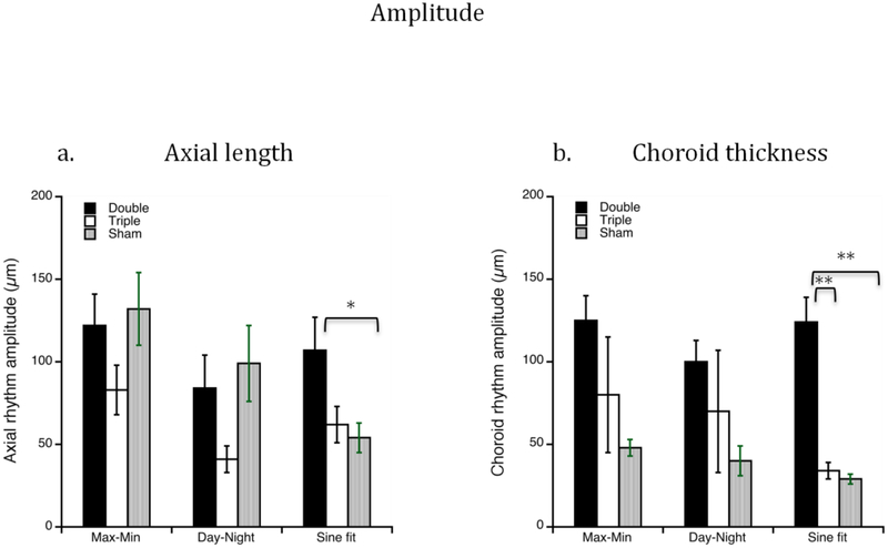
In chicks, axial length and choroidal thickness undergo circadian oscillations. The choroid is innervated by both branches of the autonomic nervous system, but their contribution(s) to these rhythms is unknown. We used two combination lesions to test this. For parasympathectomy, nerve VII was sectioned presynaptic to the pterygopalatine ganglia, and the ciliary post-ganglionics were cut (double lesion; n = 8). Triple lesions excised the sympathetic superior cervical ganglion as well (n = 8). Sham surgery was done in controls (n = 7). 8-14 days later, axial dimensions were measured with ultrasonography at 4-h intervals over 24 h. Rhythm parameters were assessed using a "best fit" function, and growth rates measured. Both types of lesions resulted in ultradian (> 1 cycle/24 h) rhythms in choroidal thickness and axial length, and increased vitreous chamber growth (Exp-fellow: double: 69 µm; triple: 104 µm; p < 0.05). For double lesions, the frequency was 1.5 cycles/day for both rhythms; for triples the choroidal rhythm was 1.5 cycles/day, and the axial was 3 cycles/day. For double lesions, the amplitudes of both rhythms were larger than those of sham surgery controls (axial: 107 vs 54 µm; choroid: 124 vs 29 µm, p < 0.05). These findings provide evidence for the involvement of abnormal ocular rhythms in the growth stimulation underlying myopia development.

Keywords: Ciliary ganglion; Myopia; Pterygopalatine; Superior cervical ganglion; Ultradian.


Figures

Figure 1.
Figure 1.
Changes in ocular dimensions. a. Interocular (experimental minus control; X-C) differences in growth rates (change per 24 hours) for vitreous chamber depth (black bars) and axial length (white bars) for double and triple lesions. b. Interocular differences (X-C) in growth rates of the anterior chamber for double and triple lesions. Double lesions caused a decrease in anterior chamber growth relative to that of fellow eyes. c. Choroidal thickness for experimental (black bars) and fellow (white bars) eyes at the end of the experiment. Error bars are standard errors of the mean. Asterisks denote statistical significance (p<0.05; paired t-tests) in the interocular differences.
Figure 2.
Figure 2.
Mean rhythms in axial length (red) and choroidal thickness (blue) for double lesions (parasympathectomy) (a) and triple lesions (b) measured at 4-hr intervals. (c) Sham surgery controls measured at 4-hr intevals. The dashed lines show the automated best fit by the Igor program. Time is on the x axis. For double lesions, both rhythms have a mean period of 16 hours (1.5 cycles/day). For triple lesions, the choroidal rhythm period is 16 hours (1.5 cycles/day), and the axial rhythm is 8 hours (3 cycles/day). Error bars are standard errors of the mean.
Figure 3.
Figure 3.
Changes over the 4-hour intervals for axial length (top, a) and choroid thickness (top, b) in the double (black bars) and triple (white bars) lesions. For axial length, note the difference in oscillations between the two lesion conditions: the triple lesions show 3 cycles, and the double lesions show 2 cycles. Bottom panels for both parameters show the sine wave fits from figure 2 for comparison. Error bars are standard errors of the mean.
Figure 4.
Figure 4.
Changes over the 8-hour intervals of day (white bars) and night (black bars) for axial length (a) and choroid thickness (b) for single lesions (PPGX and CGX), double and triple lesions. The fellow eyes (all experimental groups combined) are also included at far right. For all lesion conditions, note the lack of diurnal (day vs night) differences for both rhythms, reflecting the increase in cycle frequency. Error bars are standard errors of the mean.
Figure 5.
Figure 5.
Mean rhythms in anterior chamber depth for double lesions (blue line), triple lesions (red line) and sham surgery controls (green line). The dotted lines are the “automated best fit” by the Igor program. Time is on the x axis. Note that the double lesion data (blue) cannot be fit to a sine function. Error bars are standard errors of the mean.
Figure 6.
Figure 6.
Amplitude derived from 3 analyses (Methods) for double lesions (black bars), triple lesions (white bars) and sham surgery controls (grey bars), for axial length (a) and choroid thickness (b). Note that double lesions result in an increase in amplitude for both axial length (a) and choroid thickness (b) using the sine wave fit analysis. Error bars are standard errors of the mean. *p<0.05; **p<0.00005
Figure 7.
Figure 7.
Rhythms in axial length (red) and choroid thickness (blue) in fellow eyes for double lesions (a) and triple lesions (b). The dotted lines are the “automated best fit” by the Igor program. Time is on the x axis. Note that rhythms are abnormal, indicating a yoking by the contralateral experimental eyes for both lesions. Error bars are standard errors of the mean.

References

    1. Blessing W, Ootsuka Y (2016) Timing of activities of daily life is jaggy: How episodic ultradian changes in body and brain temperature are integrated into this process. Temperature 3: 371–383. - PMC - PubMed
    1. Bourguignon C, Storch KF (2017) Control of rest: Activity by a dopaminergic ultradian oscillator and the circadian clock. Front Neurol 8: 614. - PMC - PubMed
    1. Buijs RM, Fleur SE, Wortel J, van Heyningen C, Zuiddam L, Mettenleiter TC, Kalsbeek A, Nagai K, Niijima A (2003) The suprachiasmatic nucleus balances sympathetic and parasympathetic output to peripheral organs through separate preautonomic neurons. J Comp Neurol 464: 36–48. - PubMed
    1. Chakraborty R, Ostrin L, Nickla D, Iuvone P, Pardue M, Stone RA (2018) Circadian rhythms, refractive development, and myopia. Ophthal Physiol Opt 38: 217–245. - PMC - PubMed
    1. De Stefano M, Mugnaini E (1997) Fine structure of the choroidal coat of the avian eye: lymphatic vessels. Invest Ophthalmol Vis Sci 38: 1241–1260. - PubMed

Publication types

LinkOut - more resources