

Annulus fibrosus cell phenotypes in homeostasis and injury: implications for regenerative strategies
- PMID: 30604562
- PMCID: PMC6417974
- DOI: 10.1111/nyas.13964
Annulus fibrosus cell phenotypes in homeostasis and injury: implications for regenerative strategies
Abstract
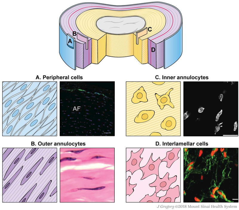
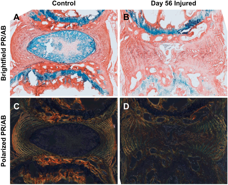
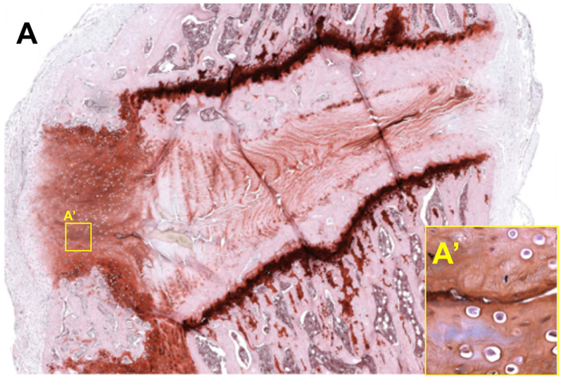
Despite considerable efforts to develop cellular, molecular, and structural repair strategies and restore intervertebral disk function after injury, the basic biology underlying intervertebral disk healing remains poorly understood. Remarkably, little is known about the origins of cell populations residing within the annulus fibrosus, or their phenotypes, heterogeneity, and roles during healing. This review focuses on recent literature highlighting the intrinsic and extrinsic cell types of the annulus fibrosus in the context of the injury and healing environment. Spatial, morphological, functional, and transcriptional signatures of annulus fibrosus cells are reviewed, including inner and outer annulus fibrosus cells, which we propose to be referred to as annulocytes. The annulus also contains peripheral cells, interlamellar cells, and potential resident stem/progenitor cells, as well as macrophages, T lymphocytes, and mast cells following injury. Phases of annulus fibrosus healing include inflammation and recruitment of immune cells, cell proliferation, granulation tissue formation, and matrix remodeling. However, annulus fibrosus healing commonly involves limited remodeling, with granulation tissues remaining, and the development of chronic inflammatory states. Identifying annulus fibrosus cell phenotypes during health, injury, and degeneration will inform reparative regeneration strategies aimed at improving annulus fibrosus healing.
Keywords: annulus fibrosus; cell phenotype; healing; intervertebral disk; intervertebral disk injury models; regeneration.
© 2018 New York Academy of Sciences.
Conflict of interest statement
Competing Interests
The authors declare no competing interests.
Figures
Similar articles
-
Mechanically tough, adhesive, self-healing hydrogel promotes annulus fibrosus repair via autologous cell recruitment and microenvironment regulation.Acta Biomater. 2024 Apr 1;178:50-67. doi: 10.1016/j.actbio.2024.02.020. Epub 2024 Feb 19. Acta Biomater. 2024. PMID: 38382832
-
Repair and Regenerative Therapies of the Annulus Fibrosus of the Intervertebral Disc.J Coll Physicians Surg Pak. 2016 Feb;26(2):138-44. J Coll Physicians Surg Pak. 2016. PMID: 26876403 Review.
-
Mechanical Factors Regulate Annulus Fibrosus (AF) Injury Repair and Remodeling: A Review.ACS Biomater Sci Eng. 2024 Jan 8;10(1):219-233. doi: 10.1021/acsbiomaterials.3c01091. Epub 2023 Dec 27. ACS Biomater Sci Eng. 2024. PMID: 38149967 Review.
-
Development of a two-step protocol for culture expansion of human annulus fibrosus cells with TGF-β1 and FGF-2.Stem Cell Res Ther. 2016 Jul 12;7(1):89. doi: 10.1186/s13287-016-0332-1. Stem Cell Res Ther. 2016. PMID: 27405858 Free PMC article.
-
Biomechanics in Annulus Fibrosus Degeneration and Regeneration.Adv Exp Med Biol. 2018;1078:409-420. doi: 10.1007/978-981-13-0950-2_21. Adv Exp Med Biol. 2018. PMID: 30357635 Review.
Cited by
-
Targeting Ferroptosis Holds Potential for Intervertebral Disc Degeneration Therapy.Cells. 2022 Nov 5;11(21):3508. doi: 10.3390/cells11213508. Cells. 2022. PMID: 36359904 Free PMC article. Review.
-
Development, Pathogenesis, and Regeneration of the Intervertebral Disc: Current and Future Insights Spanning Traditional to Omics Methods.Front Cell Dev Biol. 2022 Mar 11;10:841831. doi: 10.3389/fcell.2022.841831. eCollection 2022. Front Cell Dev Biol. 2022. PMID: 35359439 Free PMC article. Review.
-
The Use of Medical Ozone in Chronic Intervertebral Disc Degeneration Can Be an Etiological and Conservative Treatment.Int J Mol Sci. 2023 Mar 31;24(7):6538. doi: 10.3390/ijms24076538. Int J Mol Sci. 2023. PMID: 37047511 Free PMC article. Review.
-
Angiopoietin-2 Promotes Mechanical Stress-induced Extracellular Matrix Degradation in Annulus Fibrosus Via the HIF-1α/NF-κB Signaling Pathway.Orthop Surg. 2023 Sep;15(9):2410-2422. doi: 10.1111/os.13797. Epub 2023 Jul 21. Orthop Surg. 2023. PMID: 37475697 Free PMC article.
-
Nonviral overexpression of Scleraxis or Mohawk drives reprogramming of degenerate human annulus fibrosus cells from a diseased to a healthy phenotype.JOR Spine. 2023 Jun 27;6(3):e1270. doi: 10.1002/jsp2.1270. eCollection 2023 Sep. JOR Spine. 2023. PMID: 37780832 Free PMC article.
References
-
- GBD 2016 Disease and Injury Incidence and Prevalence Collaborators. Global, regional, and national incidence, prevalence, and years lived with disability for 328 diseases and injuries for 195 countries, 1990–2016: a systematic analysis for the Global Burden of Disease Study 2016. The Lancet 2017;390(10100):1211–1259. doi:10.1016/S0140-6736(17)32154-2. - DOI - PMC - PubMed
-
- Torre OM, Cruz MA, Hecht AC, Iatridis JC. Chapter 15: Annulus Fibrosus Repair. In: Härtl R, Bonassar LJ, eds. Biological Approaches to Spinal Disc Repair and Regeneration for Clinicians.; 2017.
Publication types
MeSH terms
Grants and funding
LinkOut - more resources
Full Text Sources
Medical
