

The split sign: The MRI equivalent of the bell clapper deformity
- PMID: 30604623
- PMCID: PMC6541176
- DOI: 10.1259/bjr.20180312
The split sign: The MRI equivalent of the bell clapper deformity
Abstract
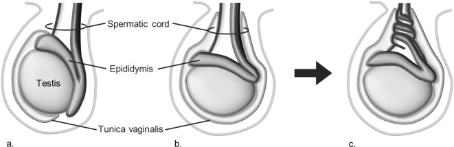
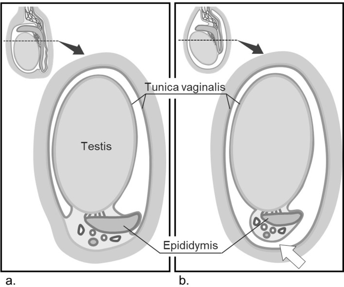
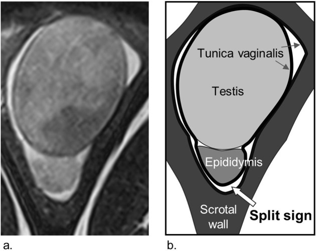
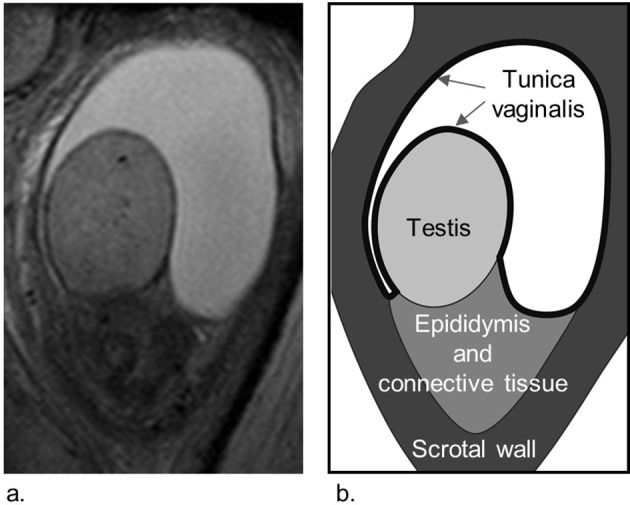
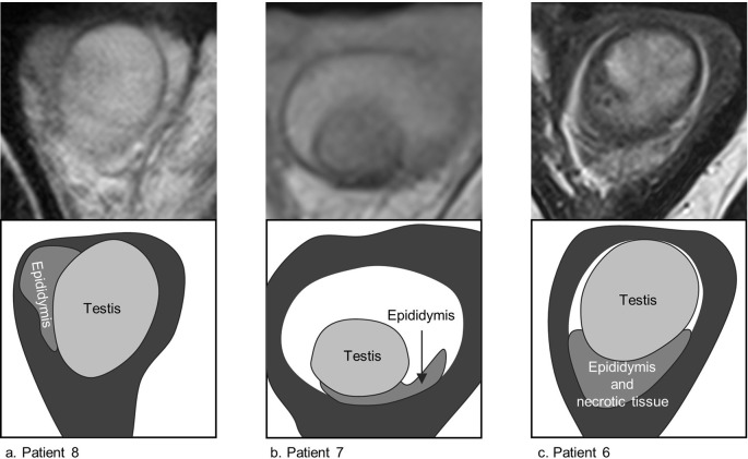
Methods:: The cases of eight patients who underwent MRI and surgery for acute scrotum between January 2010 and January 2017 were evaluated. We recorded whether hyperintense fluid on T2 weighted images existed between the posterior aspect of the epididymis and the scrotal wall ("split sign") and investigated if it correlated with BCD in surgical findings.
Results:: In one patient without hydrocele, readers were unable to evaluate the anatomy of the tunica vaginalis. Among seven patients with hydrocele, five had the split sign and all were surgically confirmed as BCD. In two patients with hydrocele but no split sign, one had normal scrotal anatomy and the other had a BCD with a necrotic testis adherent to the scrotal wall.
Conclusion:: The split sign on MRI corresponded well to the lack of fixation of the epididymis to the scrotal wall and detected BCD with high sensitivity (5/6).
Advances in knowledge:: A hyperintense area on T2 weighted image between the posterior aspect of the epididymis and scrotal wall ("split sign") is a useful MRI finding for diagnosing BCD.
Figures
Similar articles
-
Sonographic differential diagnosis of acute scrotum: real-time whirlpool sign, a key sign of torsion.J Ultrasound Med. 2006 May;25(5):563-74. doi: 10.7863/jum.2006.25.5.563. J Ultrasound Med. 2006. PMID: 16632779 Clinical Trial.
-
Scrotal pathology in pediatrics with sonographic imaging.Curr Probl Diagn Radiol. 2000 Nov-Dec;29(6):185-205. doi: 10.1016/s0363-0188(00)90013-6. Curr Probl Diagn Radiol. 2000. PMID: 11104171 Review.
-
Acute Scrotum Secondary to Torsion of a Tunica Vaginalis Cyst.Urology. 2020 Oct;144:e1-e3. doi: 10.1016/j.urology.2020.07.001. Epub 2020 Jul 16. Urology. 2020. PMID: 32683065
-
Does color Doppler sonography improve the clinical assessment of patients with acute scrotum?Eur J Radiol. 2006 Oct;60(1):120-4. doi: 10.1016/j.ejrad.2006.04.016. Epub 2006 May 30. Eur J Radiol. 2006. PMID: 16730939
-
The bell-clapper deformity of the testis: The definitive pathological anatomy.J Pediatr Surg. 2021 Aug;56(8):1405-1410. doi: 10.1016/j.jpedsurg.2020.06.023. Epub 2020 Jun 25. J Pediatr Surg. 2021. PMID: 32762939
Cited by
-
When to ask for an MRI of the scrotum.Andrology. 2021 Sep;9(5):1395-1409. doi: 10.1111/andr.13032. Epub 2021 Jun 11. Andrology. 2021. PMID: 33964115 Free PMC article. Review.
-
Testicular volume and Tanner stage: determinant factors for testicular torsion.Einstein (Sao Paulo). 2022 Apr 20;20:eAO6605. doi: 10.31744/einstein_journal/2022AO6605. eCollection 2022. Einstein (Sao Paulo). 2022. PMID: 35476083 Free PMC article.
References
-
- Shittu OB, Idowu OE, Malomo AO, Ajani RSA. Intrascrotal anomalies related to testicular torsion in nigerians: an anatomical study. Afr J Urol 2006; 12: 24–8.
MeSH terms
LinkOut - more resources
Full Text Sources
Medical
