. 2019 Mar 1;316(3):L470-L486.
doi: 10.1152/ajplung.00304.2018. Epub 2019 Jan 3.

Cinnamaldehyde in flavored e-cigarette liquids temporarily suppresses bronchial epithelial cell ciliary motility by dysregulation of mitochondrial function

Affiliations

Cinnamaldehyde in flavored e-cigarette liquids temporarily suppresses bronchial epithelial cell ciliary motility by dysregulation of mitochondrial function

Phillip W Clapp et al. Am J Physiol Lung Cell Mol Physiol. .

Abstract

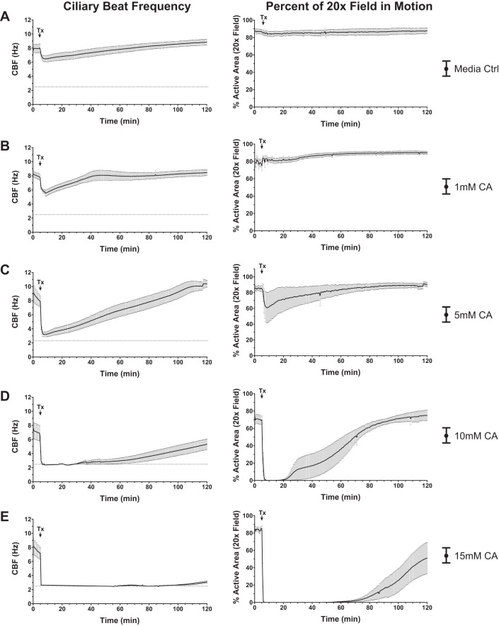
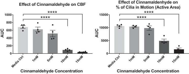
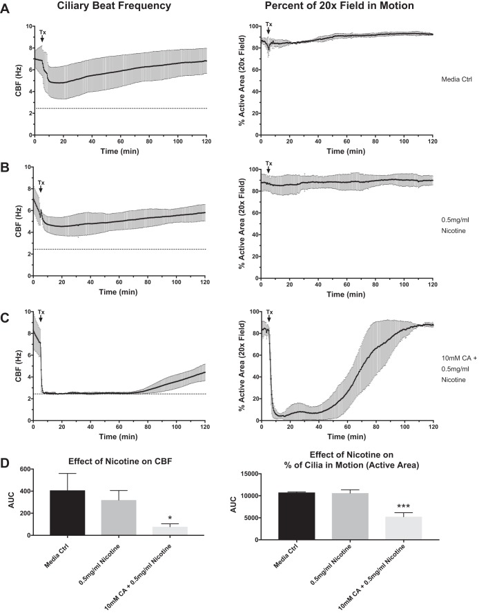
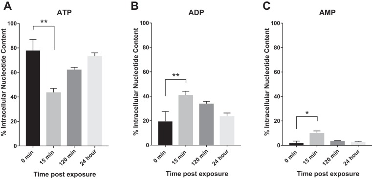
Aldehydes in cigarette smoke (CS) impair mitochondrial function and reduce ciliary beat frequency (CBF), leading to diminished mucociliary clearance (MCC). However, the effects of aldehyde e-cigarette flavorings on CBF are unknown. The purpose of this study was to investigate whether cinnamaldehyde, a flavoring agent commonly used in e-cigarettes, disrupts mitochondrial function and impairs CBF on well-differentiated human bronchial epithelial (hBE) cells. To this end, hBE cells were exposed to diluted cinnamon-flavored e-liquids and vaped aerosol and assessed for changes in CBF. hBE cells were subsequently exposed to various concentrations of cinnamaldehyde to establish a dose-response relationship for effects on CBF. Changes in mitochondrial oxidative phosphorylation and glycolysis were evaluated by Seahorse Extracellular Flux Analyzer, and adenine nucleotide levels were quantified by HPLC. Both cinnamaldehyde-containing e-liquid and vaped aerosol rapidly yet transiently suppressed CBF, and exposure to cinnamaldehyde alone recapitulated this effect. Cinnamaldehyde impaired mitochondrial respiration and glycolysis in a dose-dependent manner, and intracellular ATP levels were significantly but temporarily reduced following exposure. Addition of nicotine had no effect on the cinnamaldehyde-induced suppression of CBF or mitochondrial function. These data indicate that cinnamaldehyde rapidly disrupts mitochondrial function, inhibits bioenergetic processes, and reduces ATP levels, which correlates with impaired CBF. Because normal ciliary motility and MCC are essential respiratory defenses, inhalation of cinnamaldehyde may increase the risk of respiratory infections in e-cigarette users.

Keywords: aldehyde; e-liquid; electronic cigarette; flavoring; toxicity; vaping.


Conflict of interest statement

No conflicts of interest, financial or otherwise, are declared by the authors.

Figures

Fig. 1.
Fig. 1.
Effects of cinnamaldehyde-containing e-liquids on airway cilia motility. AD: following 5-min baseline measurements, well-differentiated primary human bronchial epithelial (hBE) cells were exposed to 1% dilutions of e-liquids (vol/vol) in cell culture medium and ciliary beat frequency (CBF; left), and percent active area (%AA) of the ×20 field of view in motion (right) was recorded for 120 min (n = 4). E: 5-min baseline measurements were recorded before well-differentiated primary hBE cells were exposed to vaped Sinicide e-liquid aerosols for 5 min. CBF and %AA were recorded for 50 min following exposures (n = 3). Data are presented as means ± SE. HCC, Hot Cinnamon Candies; PG/VG, propylene glycol/vegetable glycerin.
Fig. 2.
Fig. 2.
Quantification of e-liquid-induced effects on cilia motility. Area under the curve (AUC) was calculated for both ciliary beat frequency (CBF) and percent active area (%AA) data collected from e-liquid exposure experiments (N = 4; A) and vaped Sinicide e-liquid experiments (n = 3; B). Significant differences from the propylene glycol/vegetable glycerin (PG/VG) control were determined by 1-way ANOVA and Holm-Sidak multiple-comparisons test. For vaped Sinicide experiments, significant differences from the PG/VG control were determined by unpaired t-test. Data presented as means ± SE. Significance represented as **P < 0.01; ●, individual data points derived from the different human bronchial epithelial cell cultures. HCC, Hot Cinnamon Candies.
Fig. 3.
Fig. 3.
Effects of cinnamaldehyde (CA) on airway cilia motility. Following 5-min baseline measurements, well-differentiated primary human bronchial epithelial (hBE) cells were exposed to either vehicle (cell culture medium) control (A) or 1, 5, 10, or 15 mM cinnamaldehyde (BE). Ciliary beat frequency (CBF; left) and percent of the ×20 field of view in motion [percent active area (%AA); right] were recorded for 120 min (n = 4). Data presented as means ± SE.
Fig. 4.
Fig. 4.
Quantification of cinnamaldehyde-induced effects on cilia motility. Area under the curve (AUC) was calculated for both ciliary beat frequency (CBF) and percent active area (%AA) data collected from each independent experiment (n = 4). Significant differences from the vehicle (cell culture medium) control were determined by 1-way ANOVA and Holm-Sidak multiple-comparisons test. Data presented as means ± SE. Significance represented as ****P < 0.0001; ●, individual data points derived from the different human bronchial epithelial cell cultures.
Fig. 5.
Fig. 5.
Ciliary beat frequency (CBF) in response to nicotine alone and cinnamaldehyde (CA) containing nicotine. Following 5-min baseline measurements, differentiated primary human bronchial epithelial (hBE) cells were exposed to vehicle (cell culture medium) control (A), 0.5 mg/ml nicotine (B), or a mixture of 10 mM cinnamaldehyde and 0.5 mg/ml nicotine (C). CBF (left) and percent of the ×20 field of view in motion [percent active area (%AA); right] were recorded for 120 min (n = 4). Data presented as means ± SE. D: area under the curve (AUC) was calculated for both CBF and %AA data collected from each independent experiment (n = 4). Significant differences from the vehicle (cell culture medium) control were determined by 1-way ANOVA and Holm-Sidak multiple comparisons test. Data presented as means ± SE. Significance represented as *P < 0.05 and ***P < 0.001.
Fig. 6.
Fig. 6.
Cinnamaldehyde (CA) dysregulates mitochondrial membrane potential in bronchial epithelial cells. A: Beas-2B cells (SV40 virus-transformed bronchial epithelial cell line) labeled with 2.0 μg/ml JC-1 were exposed to vehicle (cell culture medium) and 0.5, 1.0, and 5.0 mM CA for 15 min. Mitochondrial depolarization was determined by a reduction in the J-aggregate (red) to JC-1 monomer (green) ratio using a fluorescence plate reader (n = 4). B: CA-induced changes in the fluorescence signals were validated using microscopy (representative images shown). Significant differences from the vehicle control were determined by 1-way ANOVA and Dunnett’s multiple-comparisons test. Data presented as means ± SE. Significance represented as **P < 0.01 and ****P < 0.0001.
Fig. 7.
Fig. 7.
Cinnamaldehyde (CA) induces a concentration-dependent reduction in bronchial epithelial cell mitochondrial respiration. A: Beas-2B cells (SV40 virus-transformed bronchial epithelial cell line) were exposed to 0.05, 0.25, 0.5, and 5.0 mM concentrations of CA, and changes in mitochondrial oxygen consumption rate (OCR) were evaluated using a Seahorse bioanalyzer (n = 4). BF: exposures of 0.25, 0.5, and 5 mM CA significantly suppressed basal OCR (B), ATP production (C), reserve capacity (D), proton leak (E), and maximal respiration (F) in a concentration-dependent manner. Significant differences from the vehicle control were determined by 1-way ANOVA and Dunnett’s multiple comparisons test. Data presented as means ± SE. Significance represented as ****P < 0.0001.
Fig. 8.
Fig. 8.
Cinnamaldehyde (CA) reduces bronchial epithelial cell glycolytic function. A: Beas-2B cells (SV40 virus-transformed bronchial epithelial cell line) were exposed to 0.05, 0.25, 0.5, and 5.0 mM concentrations of CA, and the rate of extracellular acidification (ECAR) was measured using a Seahorse bioanalyzer (n = 4). B and C: exposures of 0.25, 0.5, and 5 mM CA significantly suppressed basal ECAR (B), whereas 0.5 and 5 mM CA significantly reduced Beas-2B cell glycolytic capacity (C). Significant differences from the vehicle control were determined by 1-way ANOVA and Dunnett’s multiple-comparisons test. Data presented as means ± SE. Significance represented as **P < 0.01 and ****P < 0.0001. Tx, treatment/challenge; Rot/Ant A, rotenone/antimycin A.
Fig. 9.
Fig. 9.
Effects of cinnamaldehyde and nicotine on mitochondrial membrane potential in differentiated primary human bronchial epithelial (Hbe) cells. Differentiated hBE cells labeled with 2.0 μg/ml JC-1 were exposed to 10.0 mM cinnamaldehyde (A), 15 mM cinnamaldehyde (B), 0.5 mg/ml nicotine (C), or 5.0 mg/ml nicotine (D) for 120 min. Mitochondrial depolarization was determined at baseline, 15 min postexposure, and 120 min postexposure by a reduction in the J-aggregate (red) to JC-1 monomer (green) ratio using a fluorescence plate reader (n = 4). Significant differences from the vehicle control were determined by 1-way ANOVA and Dunnett’s multiple comparisons test. Data presented as means ± SE. Significance represented as *P < 0.05.
Fig. 10.
Fig. 10.
Exposure of well-differentiated human bronchial epithelial (hBE) cells to cinnamaldehyde causes a rapid but temporary reduction in intracellular ATP levels. Well-differentiated hBE cells were exposed to 10 mM cinnamaldehyde for 15 min, 120 min, and 24 h, and the effect on intracellular adenosine nucleotides was quantified by high-performance liquid chromatography (n = 4). Exposure caused a significant reduction in ATP levels after 15 min (A) that correlated with increased levels of ADP (B) and AMP (C). Significant differences from the vehicle control were determined by 1-way ANOVA and Dunnett’s multiple-comparisons test. Data presented as means ± SE. Significance represented as *P < 0.05 and **P < 0.01.
Fig. 11.
Fig. 11.
Chemical inhibition of well-differentiated human bronchial epithelial (hBE) cell glycolytic function and mitochondrial respiration significantly alters cilia motility. A and B: inhibition of glycolytic function with 2-deoxy-d-glucose (2-DG) significantly increased ciliary beat frequency (CBF; A), whereas inhibition of mitochondrial respiration with antimycin A caused a significant reduction in CBF and percent active areas (%AA) (B). C: addition of both chemical inhibitors produced a greater reduction in CBF and %AA. However, ciliary motility was restored by removing inhibitors and supplementing cultures with fresh growth medium (n = 4). Significant differences from baseline CBF and %AA were determined by 1-way ANOVA and Holm-Sidak multiple-comparisons test. Data presented as means ± SE. Significance represented as *P < 0.05, **P < 0.01, and ***P < 0.001.

Similar articles

Cited by

References

    1. Bahl V, Lin S, Xu N, Davis B, Wang YH, Talbot P. Comparison of electronic cigarette refill fluid cytotoxicity using embryonic and adult models. Reprod Toxicol 34: 529–537, 2012. doi:10.1016/j.reprotox.2012.08.001. - DOI - PubMed
    1. Barnes PJ, Burney PG, Silverman EK, Celli BR, Vestbo J, Wedzicha JA, Wouters EF. Chronic obstructive pulmonary disease. Nat Rev Dis Primers 1: 15076, 2015. doi:10.1038/nrdp.2015.76. - DOI - PubMed
    1. Barrington-Trimis JL, Samet JM, McConnell R. Flavorings in electronic cigarettes: an unrecognized respiratory health hazard? JAMA 312: 2493–2494, 2014. doi:10.1001/jama.2014.14830. - DOI - PMC - PubMed
    1. Behar RZ, Davis B, Wang Y, Bahl V, Lin S, Talbot P. Identification of toxicants in cinnamon-flavored electronic cigarette refill fluids. Toxicol In Vitro 28: 198–208, 2014. doi:10.1016/j.tiv.2013.10.006. - DOI - PubMed
    1. Behar RZ, Luo W, Lin SC, Wang Y, Valle J, Pankow JF, Talbot P. Distribution, quantification and toxicity of cinnamaldehyde in electronic cigarette refill fluids and aerosols. Tob Control 25, Suppl 2: ii94–ii102, 2016. doi:10.1136/tobaccocontrol-2016-053224. - DOI - PMC - PubMed

Publication types

MeSH terms