Review
. 2019 Mar;92(1095):20180768.
doi: 10.1259/bjr.20180768. Epub 2019 Jan 3.

Imaging in myeloma with focus on advanced imaging techniques

Affiliations
Review

Imaging in myeloma with focus on advanced imaging techniques

Tara Barwick et al. Br J Radiol. 2019 Mar.

Abstract

In recent years, there have been major advances in the imaging of myeloma with whole body MRI incorporating diffusion-weighted imaging, emerging as the most sensitive modality. Imaging is now a key component in the work-up of patients with a suspected diagnosis of myeloma. The International Myeloma Working Group now specifies that more than one focal lesion on MRI or lytic lesion on whole body low-dose CT or fludeoxyglucose (FDG) PET/CT fulfil the criteria for bone damage requiring therapy. The recent National Institute for Health and Care Excellence myeloma guidelines recommend imaging in all patients with suspected myeloma. In addition, there is emerging data supporting the use of functional imaging techniques (WB-DW MRI and FDG PET/CT) to predict outcome and evaluate response to therapy. This review summarises the imaging modalities used in myeloma, the latest guidelines relevant to imaging and future directions.


Figures
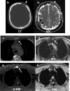
Figure 1.
Figure 1.
Limitation of WBLDCT: A 58-year-old male staging WBLDCT (a) detected lytic left frontal lesion only (white arrowheads). WB-MRI (b, d, e, f) detected additional active extra osseous dural disease (black arrowheads) and osseous disease in sternum (white arrows), high signal in the water only DIXON sequence (d), high signal on the b900 (e) with restricted diffusion on the ADC map (f). This was occult on CT (c). ADC, apparent diffusion coefficient; WBLDCT, whole body low dose CT.
Figure 2.
Figure 2.
Staging WB-MRI: A 65-year-old with non-secretory myeloma staging WB-MRI. The inverse grey scale b900 MIP (a), axial water only DIXON (b, c), axial b900 (d, e) and axial ADC (f, g) show multiple active lesions (arrows & asterisks). ADC, apparent diffusion coefficient; MIP, maximum intensity projection.
Figure 3.
Figure 3.
Active vs treated WB-MRI: A 49-year-old with suspected relapse post autologous SCT. Restaging WB-MRI (a, b) shows mixed active (asterisks) and treated disease (white arrowheads). WBLDCT (c) cannot distinguish treated vs active disease (white arrows). WBLDCT, whole body low dose CT; WB-MRI, whole body MRI.
Figure 4.
Figure 4.
FDG PET/CT Active vs treated disease: A 61-year-old with suspected relapse post autologous stem cell transplant. FDG PET/CT MIP (a) shows multiple lesions (black arrows). On the CT (b, c) and fused (d, e), there is a mixture of active (white arrowheads) and treated disease lytic on CT but not avid (white arrows). FDG, fludeoxyglucose; MIP, maximum intensity projection.
Figure 5.
Figure 5.
Suspected solitary plasmacytoma: A 75-year-old presenting with pathological fracture of the right mid-clavicle, presumed to be a solitary plasmacytoma on standard skeletal survey (a, black arrow). FDG PET/CT (b, c, d, e, f) detected additional lesions in the thoracic spine lytic on CT and left femur, occult on CT (white arrows). FDG, fludeoxyglucose, PET, positron emission tomography.

References

    1. CRUK myeloma statistics . 2018. . Available from: https://www.cancerresearchuk.org/health-professional/cancer-statistics/s... .
    1. Rajkumar SV , Dimopoulos MA , Palumbo A , Blade J , Merlini G , Mateos MV , et al. . International Myeloma Working Group updated criteria for the diagnosis of multiple myeloma . Lancet Oncol 2014. ; 15 : e538 – e548 . doi: 10.1016/S1470-2045(14)70442-5 - DOI - PubMed
    1. Kumar S , Paiva B , Anderson KC , Durie B , Landgren O , Moreau P , et al. . International Myeloma Working Group consensus criteria for response and minimal residual disease assessment in multiple myeloma . Lancet Oncol 2016. ; 17 : e328 – e346 . doi: 10.1016/S1470-2045(16)30206-6 - DOI - PubMed
    1. CRUK myeloma survival statistics . 2018. . Available from: https://www.cancerresearchuk.org/health-professional/cancer-statistics/s... .
    1. Myeloma: diagnosis and management. NICE guideline [NG35] . 2016. . Available from: https://www.nice.org.uk/guidance/ng35 .

MeSH terms

Substances