. 2019 Jan 3;10(1):45.
doi: 10.1038/s41467-018-07845-z.

Genome editing in primary cells and in vivo using viral-derived Nanoblades loaded with Cas9-sgRNA ribonucleoproteins

Affiliations

Genome editing in primary cells and in vivo using viral-derived Nanoblades loaded with Cas9-sgRNA ribonucleoproteins

Philippe E Mangeot et al. Nat Commun. .

Abstract

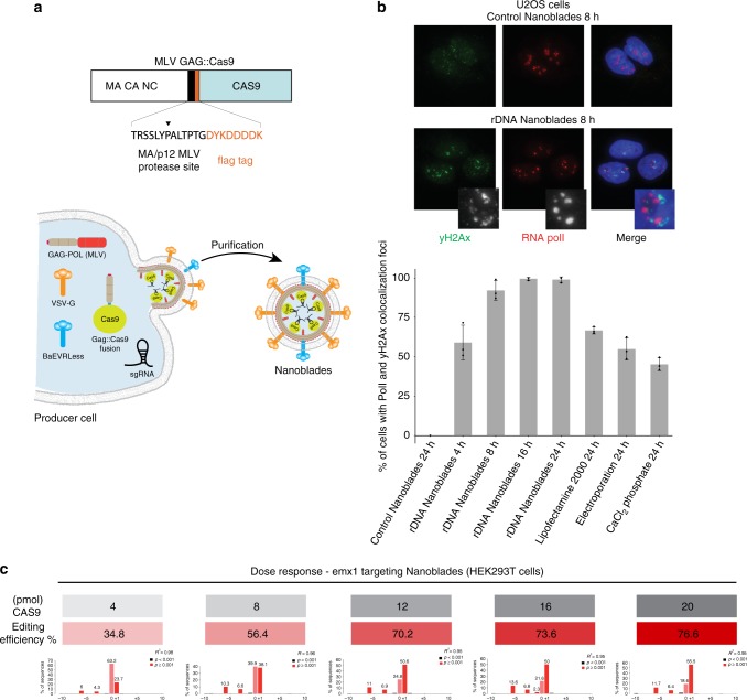
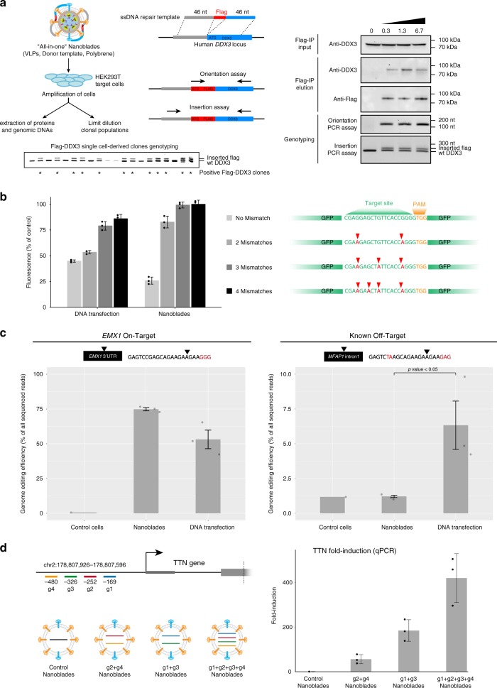
Programmable nucleases have enabled rapid and accessible genome engineering in eukaryotic cells and living organisms. However, their delivery into target cells can be technically challenging when working with primary cells or in vivo. Here, we use engineered murine leukemia virus-like particles loaded with Cas9-sgRNA ribonucleoproteins (Nanoblades) to induce efficient genome-editing in cell lines and primary cells including human induced pluripotent stem cells, human hematopoietic stem cells and mouse bone-marrow cells. Transgene-free Nanoblades are also capable of in vivo genome-editing in mouse embryos and in the liver of injected mice. Nanoblades can be complexed with donor DNA for "all-in-one" homology-directed repair or programmed with modified Cas9 variants to mediate transcriptional up-regulation of target genes. Nanoblades preparation process is simple, relatively inexpensive and can be easily implemented in any laboratory equipped for cellular biology.


Conflict of interest statement

P.E.M., T.O., and E.P.R. are named as inventors on a patent relating to the Nanoblades technology (patent applicants: Institut National de la Sante et de la Recherche Medicale (INSERM), Centre National de la Recherche Scientifique (CNRS), Ecole Normale Superieure de Lyon, Universite Claude Bernard Lyon 1, Villeurb-Anne Cedex; name of inventors: Theophile Ohlmann, Mathieu Misery, Philippe Mangeot, Emiliano Ricci; application number: WO 2017/068077 Al; patent status: published, 27th April 2017; all aspects of the manuscript are covered by the patent application. The remaining authors declare no competing interests.

Figures

Fig. 1
Fig. 1
Nanoblade-mediated genome editing. a Scheme describing the MLV Gag::Cas9 fusion and the Nanoblade production protocol based on the transfection of HEK-293T cells by plasmids coding for Gag-Pol, Gag::Cas9, VSV-G, BaEVRLess, and the sgRNA. b Top panel, immunofluorescence analysis of γ-H2AX (green), RNA polI (red) in U2OS cells 8 h after being transduced with control Nanoblades or with Nanoblades targeting ribosomal DNA genes. Bottom panel, quantification of γ-H2AX and RNA polI colocalization foci in U2OS cells at different times after Nanoblades transduction or after classical DNA transfection methods (n = 3, error bars correspond to standard deviation). c Dose response of Nanoblades. HEK-293T cells were transduced with increasing amounts of Nanoblades targeting human EMX1 (n = 1 displayed). The exact amount of Cas9 used for transduction was measured by dot blot (in gray). Genome editing was assessed by Sanger sequencing and Tide analysis (in red)
Fig. 2
Fig. 2
Genome editing in primary cells transduced with Nanoblades. a Left panel, editing efficiency at the EMX1 locus (measured by high-throughput sequencing on the Illumina Miseq platform) of human-induced pluripotent stem cells (hiPSCs) transduced with Nanoblades targeting human EMX1 (n = 3). Right panel, expression of pluripotency markers measured by qPCR in control cells and cells transduced with Nanoblades targeting EMX1 (n = 3). b Left and middle panels, fluorescence microscopy and FACS analysis of GFP expressing BMDMs transduced at the bone marrow stage (day 0 after bone marrow collection) with control Nanoblades or Nanoblades targeting the GFP-coding sequence (n = 3). Right top panel, T7 endonuclease assay against the GFP sequence from Nanoblades-treated BMDMs. Right bottom panel, cytokine expression levels (measured by qPCR) in untreated or Nanoblade-treated cells upon LPS stimulation (n = 4). c T7 endonuclease assay against mouse Fto or human EMX1 genomic sequences amplified by PCR from primary mouse bone marrow cells transduced with Nanoblades or electroporated with recombinant Cas9-sgRNA RNPs. For bone marrow cells, two electroporation settings were tested. Lanes numbered #1–#3 correspond to biological replicates. Editing efficiencies were calculated by TIDE analysis of the Sanger sequencing electropherograms for each PCR amplicon d Left panel, excision of a 160 bp DNA fragment of MYD88 using Nanoblades. Middle panel PCR results obtained in human primary hepatocytes transduced with Nanoblades. Right-panel (top), FACS analysis of CD34+ cells purified from human cord-blood. Bottom, genome editing at the MYD88 locus assessed by PCR in untreated and Nanoblades-treated CD34+ cells. Error bars in all figures correspond to standard deviation
Fig. 3
Fig. 3
“All-in-one” Nanoblades for knock-in experiments and assessment of Nanoblades off-target activity. a Left panel, Nanoblades targeting human DDX3 close to its start codon were complexed with a donor ssDNA bearing homology arms to the targeted locus and a Flag-tag sequence in the presence of polybrene. HEK293T cells were then transduced with these “All-in-one” Nanoblades. After cell amplification, a fraction of cells were collected to extract genomic DNA and total proteins while the remaining cells were cultured to obtain single-cell clonal populations. Right panel, insertion of the Flag-tag in HEK-293T cells transduced with “all-in-one” Nanoblades complexed with increasing amounts of donor ssDNA was assessed by Flag-immunoprecipitation followed by western-blot using anti-flag or anti-DDX3 antibodies in the input and Flag-immunoprecipitation elution fractions. Flag insertion was also assessed by PCR using a forward primer in the flag-sequence and a reverse primer in the DDX3 locus (Orientation PCR assay) or using primers flanking the Flag sequence (Insertion PCR assay). Bottom panel, Flag-insertion in 20 different single-cell-derived clones was assessed by PCR using primers flanking the Flag-sequence. b Left panel, off-target monitoring in immortalized mouse macrophages stably expressing GFP transgenes bearing silent mutations in the region targeted by the sgRNA. Right panel, cells were transfected with plasmids coding for Cas9 and the sgRNA or transduced with Nanoblades. GFP expression was measured by FACS 72 h after transfection/transduction (n = 3). c Left and right panels, gene-editing at the EMX1 on-target site and the MFAP1 intronic off-target site measured by high-throughput sequencing in untreated cells (control cells) and cells transduced with EMX1 Nanoblades (Nanoblades) or transfected with plasmids coding for Cas9 and the EMX1 sgRNA (DNA transfection) (n = 3). Statistical significance of the Nanoblades and DNA transfection comparison at the on-target site was computed using a two-tail Student test. d Left panel, position of sgRNAs targeting the promoter of TTN and VLPs with different combination of sgRNAs produced for the experiment. Right-panel, TTN mRNA expression levels (normalized to Control) as measured by qPCR in MCF7 transduced with VLPs (n = 3). Error bars in all figures correspond to standard deviation
Fig. 4
Fig. 4
Generation of transgenic mice using Nanoblades. a Left panel, scheme describing injection of mCherry VLPs or Nanoblades in the perivitelline space of mouse 1-cell embryos. Right panel, fluorescence microscopy of mouse blastocysts injected with mCherry VLPs at the single-cell stage. b Scheme of the design strategy to target the mouse Tyr locus (adapted from ref. ). Upon editing and NHEJ repair, the HinfI restriction site becomes inactive. c Survival rates of injected embryos at two-cell, blastocyst, and newborn stage (the latter obtained from experiments presented in Supplementary figure 5). d T7 endonuclease (top panel) and HinfI restrictions (bottom panel) assays on PCR fragments amplified from the Tyr locus of Control or Nanoblades-injected embryos. e Top left panel, photographs of F0 mice generated from embryos injected with Nanoblades programmed with two sgRNAs targeting the Tyr locus. Top-right panel, phenotype, editing efficiency (as measured by TIDE analysis of the Sanger-sequencing electropherograms) and the main INDEL type as detected by Sanger sequencing of individual PCR clones. Bottom-panel, alignment of individual PCR clones obtained from the Tyr locus of F0 mice against the mouse mm10 genome indicating the main observed INDELs in chimeric mice (mouse #4, #7, and #8) and total excision of the Tyr sequence between the sgRNA1 and sgRNA2 targeting loci for the complete albino mouse (mouse #3). The Sanger sequencing electropherogram from the bulk PCR amplicon obtained from mouse #3 indicates complete editing at both targeted sites
Fig. 5
Fig. 5
Inactivation of Hpd in the liver of tyrosinaemic FRG mice. a Scheme of the experimental approach to target the liver of FRG mice. b T7 endonuclease assay to monitor genome editing at the Hpd gene in immortalized mouse macrophages and in the liver or spleen of injected mice. Samples were quantified using a Tapestation chip

References

    1. Jinek M, et al. A programmable dual-RNA-guided DNA endonuclease in adaptive bacterial immunity. Sci. N. Y. NY. 2012;337:816–821. doi: 10.1126/science.1225829. - DOI - PMC - PubMed
    1. Gheysen D, Jacobs E, de Foresta F, Thiriart C. Assembly and release of HIV-1 precursor Pr55gag virus-like particles from recombinant baculovirus-infected insect cells. Cell. 1989;59:103–112. doi: 10.1016/0092-8674(89)90873-8. - DOI - PubMed
    1. Kaczmarczyk SJ, Sitaraman K, Young HA, Hughes SH, Chatterjee DK. Protein delivery using engineered virus-like particles. Proc. Natl Acad. Sci. USA. 2011;108:16998–17003. doi: 10.1073/pnas.1101874108. - DOI - PMC - PubMed
    1. Voelkel C, et al. Protein transduction from retroviral Gag precursors. Proc. Natl Acad. Sci. USA. 2010;107:7805–7810. doi: 10.1073/pnas.0914517107. - DOI - PMC - PubMed
    1. O’connor, T. E., Rauscher, F. J. & Zeigel, R. F. Density gradient centrifugation of a murine leukemia virus. Sci. N. Y. NY144, 1144–1147 (1964). - PubMed

Publication types

MeSH terms

Substances