. 2019 Jan 3;10(1):27.
doi: 10.1038/s41467-018-07861-z.

A switch in the poly(dC)/RmlB complex regulates bacterial persister formation

Affiliations

A switch in the poly(dC)/RmlB complex regulates bacterial persister formation

Xu Chen et al. Nat Commun. .

Retraction in

Abstract

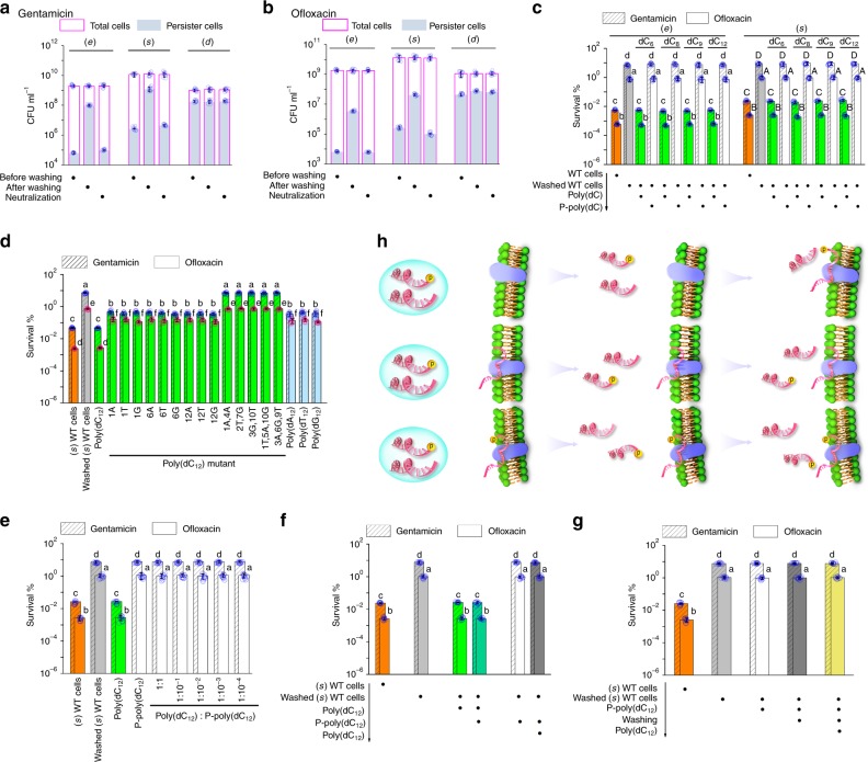
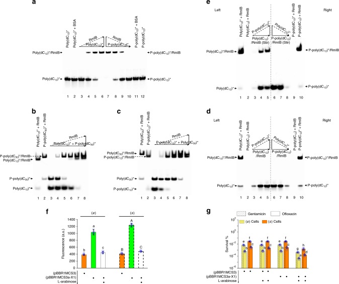
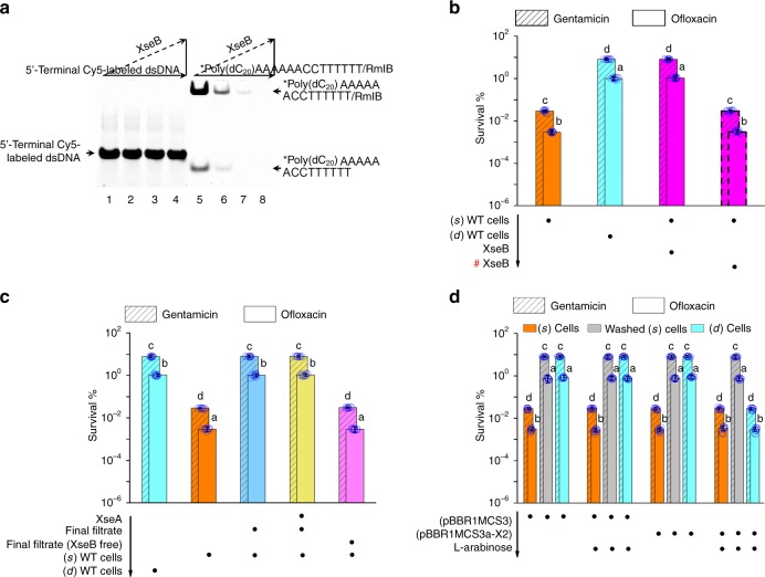
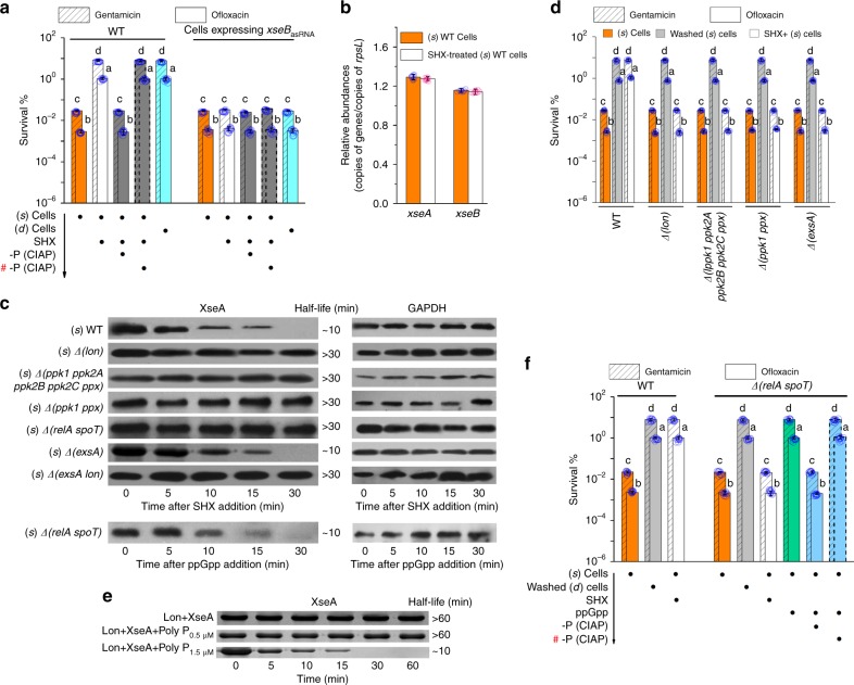
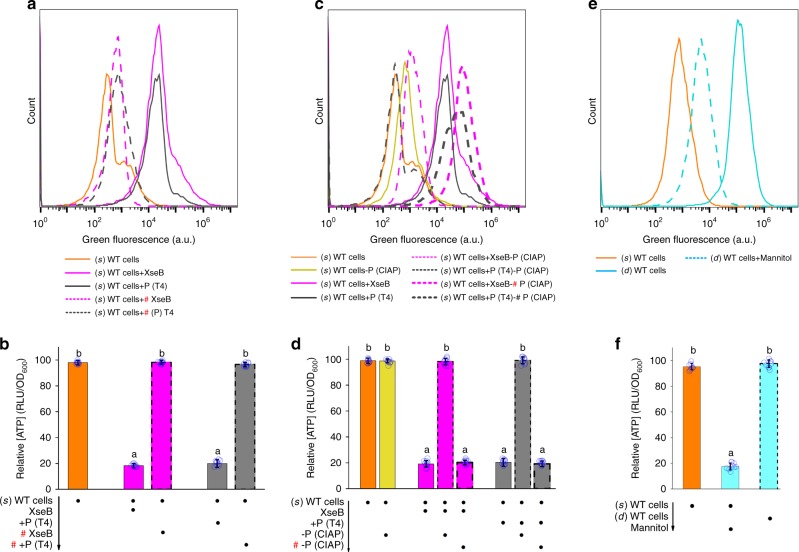
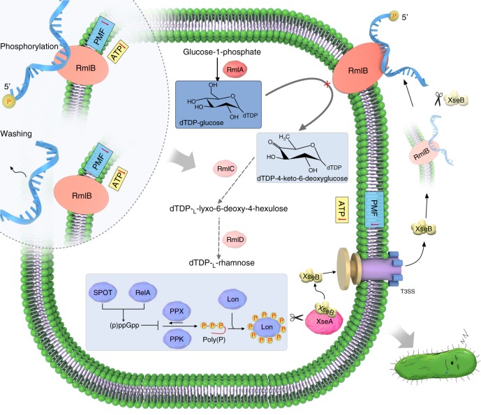
Bacterial persisters are phenotypic variants that tolerate exposure to lethal antibiotics. These dormant cells are responsible for chronic and recurrent infections. Multiple mechanisms have been linked to persister formation. Here, we report that a complex, consisting of an extracellular poly(dC) and its membrane-associated binding protein RmlB, appears to be associated with persistence of the opportunistic pathogen Pseudomonas aeruginosa. Environmental stimuli triggers a switch in the complex physiological state (from poly(dC)/RmlB to P-poly(dC)/RmlB or RmlB). In response to the switch, bacteria decrease proton motive force and intracellular ATP levels, forming dormant cells. This alteration in complex status is linked to a (p)ppGpp-controlled signaling pathway that includes inorganic polyphosphate, Lon protease, exonuclease VII (XseA/XseB), and the type III secretion system. The persistence might be also an adaptive response to the lethal action of the dTDP-L-rhamnose pathway shutdown, which occurs due to switching of poly(dC)/RmlB.


Conflict of interest statement

The authors declare no competing interests.

Figures

Fig. 1
Fig. 1
Extracellular poly(dC) modulates PAO1 persistence. a, b The number of total and persister cells before and after washing (e), (s), or (d) WT cells as well as after resuspending corresponding washed cells in their native supernatants (neutralization). c Persistence in washed (e) WT cells or washed (s) WT cells plus short poly(dC) or P-poly(dC). Poly(dC) instead of P-poly(dC) reduced persistence in washed (e) WT cells and washed (s) WT cells. d Persistence in washed (s) WT cells plus each poly(dC12) mutant, poly(dA12), poly(dT12), or poly(dG12). These ssDNA led to a nearly 10-fold reduction in persistence of washed (s) WT cells. As a control, poly(dC12) caused a more than 100-fold reduction in persistence of washed (s) WT cells. e Persistence in washed (s) WT cells plus poly(dC12) and P-poly(dC12) with different molar ratios. P-poly(dC12) abolished the regulatory activity of poly(dC12) in persistence of washed (s) WT cells. f Persistence in washed (s) WT cells plus poly(dC12) for 10 min and then P-poly(dC12) for 10 min or plus P-poly(dC12) for 10 min and then poly(dC12) for 10 min. P-poly(dC12) did not regulate persistence in poly(dC12)-pretreated washed (s) WT cells and vice versa. g Persistence in washed (s) WT cells plus P-poly(dC12) for 10 min, washing again, and then plus poly(dC12) for 10 min. These treatments did not reduce persistence in washed (s) WT cells. h Proposed model for functional antagonism between poly(dC) and P-poly(dC). All experiments were performed in biological triplicates. Error bars represent standard deviations. Dot plots overlaid on bar graphs represent individual data points. Black spot, treatment step or object; arrow, treatment order
Fig. 2
Fig. 2
RmlB controls persistence in PAO1. a Formation of DNA/protein complex by RmlB binding poly(dC12)* or P-poly(dC12)*. In lanes 3–6, a constant concentration of poly(dC12)* and an increasing concentration of His-tagged RmlB were mixed together. In lanes 7–10, a constant concentration of P-poly(dC12)* and a decreasing concentration of His-tagged RmlB were mixed together. b Order of RmlB binding poly(dC12)* and P-poly(dC30)*. In lanes 3–8, a constant concentration of poly(dC12)*/P-poly(dC30)* mixture and an increasing concentration of His-tagged RmlB were mixed together. c Order of RmlB binding P-poly(dC12)* and poly(dC30)*. In lanes 3–8, a constant concentration of P-poly(dC12)*/poly(dC30)* mixture and an increasing concentration of His-tagged RmlB were mixed together. d, e Substitutability of ssDNA in poly(dC12)/RmlB and P-poly(dC12)/RmlB. In lanes 3–5, a constant concentration of poly(dC12)/RmlB and an increasing concentration of P-poly(dC12)* were mixed together with (e) or without (d) vortex. In lanes 6–8, a constant concentration of P-poly(dC12)/RmlB and a decreasing concentration of poly(dC12)* were mixed with (e) or without (d) vortex. f Red fluorescence of cells harboring pBBR1MCS3a-X1 with or without l-arabinose induction. The RmlB fused with mCherry was expressed from a native promoter of rmlB in pBBR1MCS3a-X1. It caused a 2.9-fold increase in fluorescence as compared to cells harboring pBBR1MCS3. Red fluorescence was decreased 2.47-fold upon expression of rmlBasRNA from pBBR1MCS3a-X1 with 0.2% l-arabinose induction. The basal level of fluorescence with the empty vector (pBBR1MCS3) should come from the metabolites of bacteria. a.u. arbitrary units. g Persistence in cells harboring pBBR1MCS3a-X1 with or without l-arabinose induction. Expressing rmlBasRNA with 0.2% l-arabinose induction caused a nearly 24-fold decrease in persistence of the (e) or (s) cells. All experiments were performed in biological triplicates. Error bars represent standard deviations. Dot plots overlaid on bar graphs represent individual data points. Black spot, treatment step or object; arrow, treatment order
Fig. 3
Fig. 3
Extracellular XseB in-situ tailors poly(dC) at 5′ terminus. a Assays for the single strand-specific 5′ → 3′ cleavage activity of XseB. A Cy5 fluorescent label was attached to 5′ terminus of ssDNA with a hairpin structure in its 3′ terminus (5′-*poly(dC20)AAAAAACCTTTTTT-3′) or was attached to 5′ terminus of dsDNA (5′-*GTCATTCTGAGAATAGTGTAG-3′). The recombinant XseB resulted in the disappearance of fluorescence in free and RmlB-bound ssDNA rather than dsDNA. b Persistence in (s) WT cells plus recombinant XseB. The XseB-treated (s) WT cells exhibited the same persister level as that in (d) WT cells. c Persistence in (s) WT cells that was resuspended in the final filtrate plus His-tagged XseA or in the XseB-free final filtrate. Both the final filtrate and the His-tagged XseA treated final filtrate increased persistence in (s) WT cells. But the XseB-free final filtrate failed to increase persistence in (s) WT cells. d Persistence in cells expressing xseBasRNA. Plasmid pBBR1MCS3a-X2 had a xseBasRNA expression cassette. Expression of xseBasRNA with 0.2% l-arabinose induction caused the loss of persistence enhancement in the (d) cells. As a control, the stimulation in persistence was still present in the (d) cells harboring pBBR1MCS3a-X2 without l-arabinose induction. Washing increased persistence in the (s) cells harboring pBBR1MCS3a-X2 no matter with or without l-arabinose induction. All experiments were performed in biological triplicates. Error bars represent standard deviations. Dot plots overlaid on bar graphs represent individual data points. Black spot, treatment step or object; arrow, treatment order; hash sign, inactivated protein
Fig. 4
Fig. 4
Involvement of stringent response in induced persistence. a Persistence in (s) WT cells plus SHX and then alkaline phosphatase (CIAP) or in (s) cells expressing xseBasRNA plus SHX. SHX increased persistence in (s) WT cells and CIAP neutralized the persistence enhancement. SHX failed to increase persistence in (s) cells expressing xseBasRNA. b Quantitative real-time PCR assays for expression of xseB and xseA in (s) WT cells before and after plus 1 mg ml−1 SHX. Copy numbers of genes were set in relation to copy numbers of housekeeping gene rpsL. Significant changes were not detected. c Protein involved in triggering the degradation of XseA. WT, Δ(lon), Δ(ppk1 ppk2A ppk2B ppk2C ppx), Δ(ppk1 ppx), Δ(relA spoT), Δ(exsA), and Δ(exsA lon) cells harboring pBBR1MCS3-xseA (His-tagged XseA) were grown in LB at 37 °C for 18 h ((s) phase). After addition of 1 mg ml−1 SHX for 30 min, samples were removed at the indicated times for western blot analysis. (s) Δ(relA spoT) mutant harboring pBBR1MCS3-xseA was incubated with 5 μg ml−1 pp(G)pp at 37 °C for 30 min. Samples were then removed at the indicated times for western blot analysis. GAPDH was used as an internal reference. d Persistence in washed (s) mutants or SHX-treated (s) mutants. Both washing and SHX induction increased persistence in the (s) Δ(lon), (s) Δ(ppk1 ppk2A ppk2B ppk2C ppx), (s) Δ(ppk1 ppx), and (s) Δ(exsA) mutants, respectively. e SDS-PAGE analysis of in vitro degradation of XseA by Lon. His-tagged Lon failed to degrade His-tagged XseA in absence of polyP or in presence of low polyP (0.5 μM), but did it in presence of high polyP (1.5 μM). f Persistence in (s) Δ(relA spoT) mutant treated by washing, SHX induction, or ppGpp induction. Both washing and ppGpp induction increased persistence in the (s) Δ(relA spoT) mutant, but SHX induction did not. CIAP neutralized the ppGpp-triggered persistence enhancement. All experiments were performed in biological triplicates. Error bars represent standard deviations. Dot plots overlaid on bar graphs represent individual data points. Black spot, treatment step or object; arrow, treatment order; hash sign, inactivated protein
Fig. 5
Fig. 5
PMF and intracellular ATP levels during complex switching. a, b PMF and intracellular ATP levels of (s) WT cells plus recombinant XseB or T4 polynucleotide kinase. c, d PMF and intracellular ATP levels of XseB-treated (s) WT cells or T4 polynucleotide kinase-treated (s) WT cells, both of which were treated with CIAP. Addition of CIAP did not affect PMF of (s) WT cells. e, f PMF and intracellular ATP levels of (s) WT cells, (d) WT cells, or (d) WT cells plus d-mannitol. All experiments were performed in biological triplicates. Error bars represent standard deviations. Dot plots overlaid on bar graphs represent individual data points. Black spot, treatment step or object; arrow, treatment order; hash sign, inactivated protein
Fig. 6
Fig. 6
A proposed model explaining poly(dC)/RmlB-mediated persistence. Activation of RelA or SpoT induces a (p)ppGpp-controlled signaling pathway, which invokes Lon to degrade intracellular exonuclease VII large subunit XseA. Free exonuclease VII small subunit XseB is delivered to extracellular space by T3SS and tailors RmlB-bound poly(dC) to form P-poly(dC), which results in in-situ switch of poly(dC)/RmlB to P-poly(dC)/RmlB. On the other hand, in-situ phosphorylation or washing of PAO1 can bypass the (p)ppGpp-controlled signaling pathway and directly switch poly(dC)/RmlB to P-poly(dC)/RmlB or individual RmlB. In response to the switch in the complex physiological state, cells decrease PMF and intracellular ATP level and shutdown the dTDP-l-rhamnose pathway, forming persistence in PAO1

Similar articles

Cited by

References

    1. Lewis K. Persister cells. Annu. Rev. Microbiol. 2010;64:357–372. doi: 10.1146/annurev.micro.112408.134306. - DOI - PubMed
    1. Kint CI, Verstraeten N, Fauvart M, Michiels J. New-found fundamentals of bacterial persistence. Trends Microbiol. 2012;20:577–585. doi: 10.1016/j.tim.2012.08.009. - DOI - PubMed
    1. Helaine S, Kugelberg E. Bacterial persisters: formation, eradication, and experimental systems. Trends Microbiol. 2014;22:417–424. doi: 10.1016/j.tim.2014.03.008. - DOI - PubMed
    1. Maisonneuve E, Gerdes K. Molecular mechanisms underlying bacterial persisters. Cell. 2014;157:539–548. doi: 10.1016/j.cell.2014.02.050. - DOI - PubMed
    1. Singh R, Barry CE, 3rd, Boshoff HIM. The three RelE homologs of Mycobacterium tuberculosis have individual, drug-specific effects on bacterial antibiotic tolerance. J. Bacteriol. 2010;192:1279–1291. doi: 10.1128/JB.01285-09. - DOI - PMC - PubMed

Publication types

MeSH terms